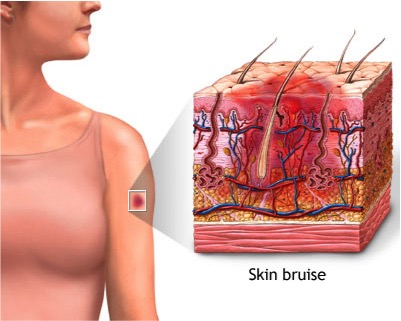
A bruise is an area of skin discoloration. A bruise occurs when small blood vessels break and leak their contents into the soft tissue beneath the skin.
A bruise occurs when a force causes blood vessels to break. Blood leaks into areas under the skin, resulting in pain, swelling, and skin discoloration.
There are three types of bruises:
Subcutaneous -- beneath the skin
Intramuscular -- within the belly of the underlying muscle
Periosteal -- bone bruise
Bruises can last from days to months, with the bone bruise being the most severe and painful.
The main symptoms are pain, swelling, and skin discoloration. The bruise begins as a pinkish red color that can be very tender to touch. It is often difficult to use the muscle that has been bruised. For example, a deep thigh bruise is painful when you walk or run.
Eventually, the bruise changes to a bluish color, then greenish-yellow, and finally returns to the normal skin color as it heals.
Place ice on the bruise to help it heal faster and to reduce swelling. Wrap the ice in a clean towel -- do not place ice directly on the skin. Apply the ice for up to 15 minutes each hour.
Keep the bruised area raised above the heart, if possible. This helps keep blood from pooling in the bruised tissue.
Try to rest the bruised body part by not overworking your muscles in that area.
If needed, take acetaminophen (Tylenol) to help reduce pain.
In the rare case of compartment syndrome, surgery is often done to relieve the extreme buildup of pressure.
Call your health care provider right away if you feel extreme pressure in a bruised part of your body, especially if the area is large or very painful. This may be due to a condition known as compartment syndrome. Increased pressure on the soft tissues and structures beneath the skin can decrease the supply of blood and oxygen to the tissues. This can be life-threatening and you should receive emergency care.
Also call your health care provider if:
You are bruising without any injury, fall, or other reason.
There are signs of infection around the bruised area including streaks of redness, pus or other drainage, or fever.
Because bruises are usually the direct result of an injury, the following are important safety recommendations:
Teach children how to be safe.
Be mindful to avoid falls around the house. For example, be careful when climbing on ladders or other objects. Avoid standing or kneeling on counter tops.
Wear seat belts in motor vehicles.
Wear proper sports equipment to pad those areas most frequently bruised, such as thigh pads, hip guards, and elbow pads in football and hockey; shin guards and knee pads in soccer and basketball.
If you have gum disease, you're not alone. Many U.S. adults currently have some form of the disease. It ranges from simple gum inflammation, called gingivitis, to serious damage to the tissue and bone supporting the teeth. In the worst cases, you can lose teeth.
In gingivitis, the gums become red and swollen. They can bleed easily. Gingivitis is a mild form of gum disease. You can usually reverse it with daily brushing and flossing and regular cleanings by a dentist or dental hygienist. Untreated gingivitis can lead to periodontitis. If you have periodontitis, the gums pull away from the teeth and form pockets that become infected. If not treated, the bones, gums and connective tissue that support the teeth are destroyed.
Domestic violence is when a person uses abusive behavior to control a partner or other family member. The abuse can be physical, emotional, economic, or sexual. It can affect people of any age, sex, culture, or class. When domestic violence is aimed at a child, it is called child abuse. Domestic violence is a crime.
Leaving an abusive relationship is not easy. You may be afraid your partner will harm you if you leave, or that you will not have the financial or emotional support you need.
Domestic violence is not your fault. You cannot stop your partner's abuse. But you can find ways to get help for yourself.
· Tell someone. The first step in getting out of an abusive relationship is often telling someone else about it. You can talk to a friend, family member, your health care provider, or a clergy member.
· Have a safety plan. This is a plan in case you need to leave a violent situation right away. Decide where you will go and what you will bring. Gather important items you will need, like credit cards, cash, or papers, in case you need to leave quickly. You can also pack a suitcase and keep it with a family member or friend.
· Get medical care. If you are hurt, get medical care from your provider or at the emergency room.
· Call the police. DO NOT hesitate to call the police if you are in danger. Domestic violence is a crime.
If a friend or family member is being abused, there are many ways you can help.
· Offer support. Your loved one may feel scared, alone, or ashamed. Let him or her know you are there to help however you can.
· DO NOT judge. Leaving an abusive relationship is difficult. Your loved one may stay in the relationship despite the abuse. Or, your loved one may leave and return many times. Try to support these choices, even if you do not agree with them.
· Help with a safety plan. Suggest that your loved one make a safety plan in case of danger. Offer your home as a safe zone if he or she needs to leave, or help find another safe place.
· Find help. Help your loved one connect with a national hotline or a domestic violence agency in your area.
Tooth loss is a process in which one or more teeth come loose and fall out. Tooth loss is normal for deciduous teeth (baby teeth), when they are replaced by a person's adult teeth. Otherwise, losing teeth is undesirable and is the result of injury or disease, such as dental avulsion, tooth decay, and gum disease. The condition of being toothless or missing one or more teeth is called edentulism.
Tooth loss typically begins around age six and continues until age twelve. The upper and lower central incisors are shed at age six to seven years. The upper and lower lateral incisors are shed at seven to eight years. The upper canines are shed at ten to twelve years. The lower canines are shed at nine to twelve years. The upper and lower first molars are shed at nine to eleven years. The upper and lower second molars are shed at ten to twelve years.[1]
As a person ages, their permanent teeth have been exposed to normal mechanical forces, such as chewing, and also more abnormal mechanical forces, such as bruxism (grinding) and traumatic injury. Permanent teeth may also be affected by oral disease.[2] There are many ways in which a person may protect his or her permanent teeth from loss.
The main method of preventing tooth loss is prevention of oral diseases. Tooth loss can be due to tooth decay and gum disease. Tooth decay is caused by increased plaque retention. Bacteria can then invade the plaque and cause dental caries (cavities). If cavities persist untreated for an extended period of time, tooth breakdown occurs.[3] Plaque retention and bacterial presence also affect the gums and bone and their ability to hold the teeth in place. Disease of the gums, known as periodontitis, leads to detachment of the supporting structures from the teeth and their eventual loss. Tooth loss due to tooth decay and gum disease may be prevented by practicing good oral hygiene, and regular check-ups at a dentist's office. Good oral hygiene consists of brushing two times a day with a fluoridated toothpaste and flossing. Dental check-ups should occur every six months. Children or adults who are incapable of caring for their own teeth should be assisted with oral hygiene in order to prevent tooth loss.[4]
In contact sports, risk of mouth trauma and tooth injury is reduced by wearing mouthguards and helmets with a facemask (e.g., a football helmet, a goalie mask).
Nightguards may also be implemented in the case of teeth grinding (bruxism) during sleep. These guards function in limiting the wear and force applied to the teeth. In turn, this minimizes the chance of loss.
In countries such as the United States, Japan, Germany, and Italy, there is a strong relationship between cigarette smoking and tooth loss. Studies have shown that an increase in exposure to cigarette smoking can increase the risk of tooth loss. In addition, studies have also found that when people stop smoking, there is a decrease in tooth loss.[5]
Proper nutrition has been shown to prevent tooth loss by providing the nutrients necessary to maintain enamel strength.[6]
Tooth loss occurs more often in people from the lower end of the socioeconomic scale.[7]
Tooth loss can occur secondary or concomitantly to many diseases. Diseases may cause periodontal disease or bone loss to prompt tooth loss. Consequently, periodontal disease may cause increased infection, which may predispose a person to other diseases. Diseases commonly related to tooth loss include, but are not limited to: cardiovascular disease,[8] cancer,[9] osteoporosis[10] and diabetes mellitus.[11] Therefore, it is important to not only maintain good oral hygiene, but also overall good health.
Maximum preservation and protection of natural teeth is best for eating and chewing; however, there are three basic ways to replace a missing tooth or teeth, including a fixed dental bridge, dentures, and dental implants. Each alternative has its own benefits and drawbacks. It is important to consider a patient's medical, financial, and emotional situation. It is recommended that a patient experiencing tooth loss visits a dentist to discuss which replacement method is best suited for his or her situation. It has been shown that a non-removable replacement, such as a bridge or implant appears to provide patients with the best sense of security and well-being.[12]
2. Jump up^ Baelum V, Luan W-M, Chen X, Fejerskov O (1997). "Predictors of tooth loss over 10 years in adult and elderly Chinese". Community Dent Oral Epidemiol. 25.
4. Jump up^ Li; et al. (2011). "Age, period and cohort analysis of regular dental care behavior and edentulism: a marginal approach.". BMC Oral Health. 11 (9).
5. Jump up^ Hanioka, T., Ojima, M., Tanaka, K., Matsuo, K., Sato, F., and Tanaka H. (2011). "Causal assessment of smoking and tooth loss:a systematic review of observational studies". BMC Public Health. 11: 221. doi:10.1186/1471-2458-11-221.
6. Jump up^ Ioannidou, E; et al. (Nov 11, 2013). "Tooth Loss Strongly Associates with Malnutrition in Chronic Kidney Disease". J Periodontol. 85 (7): 899–907. doi:10.1902/jop.2013.130347. PMID24215204.
Your kneecap (patella) sits over the front of your knee joint. As you bend or straighten your knee, the underside of the patella glides over the bones that make up the knee.
Strong tendons help attach the kneecap to the bones and muscles that surround the knee. These tendons are called:
· The patellar tendon (where the kneecap attaches to the shin bone)
· The quadriceps tendon (where the thigh muscles attach to the top of the kneecap)
Anterior knee pain begins when the kneecap does not move properly and rubs against the lower part of the thigh bone. This may occur because:
· The kneecap is in an abnormal position (also called poor alignment of the patellofemoral joint).
· There is tightness or weakness of the muscles on the front and back of your thigh.
· You are doing too much activity that places extra stress on the kneecap (such as running, jumping or twisting, skiing, or playing soccer).
· You have flat feet.
Anterior knee pain is more common in:
· People who are overweight
· People who have had a dislocation, fracture, or other injury to the kneecap
· Runners, jumpers, skiers, bicyclists, and soccer players who exercise often
· Teenagers and healthy young adults, more often girls
Other possible causes of anterior knee pain include:
· Arthritis
· Pinching of the inner lining of the knee during movement (called synovial impingement or plica syndrome)
The health care provider will perform a physical exam. The knee may be tender and mildly swollen. Also, the kneecap may not be perfectly lined up with the thigh bone (femur).
When you flex your knee, you may feel a grinding feeling below the kneecap. Pressing the kneecap when the knee is straightening out may be painful.
Your provider may want you to do a single leg squat to look at muscle imbalance and your core stability.
X-rays are very often normal. However, a special x-ray view of the kneecap may show signs of arthritis or tilting.
Resting the knee for a short period of time and taking nonsteroidal anti-inflammatory drugs (NSAIDs) such as ibuprofen, naproxen, or aspirin may help relieve pain.
Other things you can do to relieve anterior knee pain include:
· Change the way you exercise.
· Learn exercises to both strengthen and stretch the quadriceps and hamstring muscles.
· Learn exercises to strengthen your core.
· Lose weight (if you are overweight).
· Use special shoe inserts and support devices (orthotics) if you have flat feet.
· Tape your knee to realign the kneecap.
· Wear the correct running or sports shoes.
Rarely, surgery for pain behind the kneecap is needed. During the surgery:
· Kneecap cartilage that has been damaged may be removed.
· Changes may be made to the tendons to help the kneecap move more evenly.
The meniscus is a C-shaped piece of tough, rubbery cartilage that acts as a shock absorber between your shinbone and thighbone. It can be torn if you suddenly twist your knee while bearing weight on it.
A torn meniscus is one of the most common knee injuries. Any activity that causes you to forcefully twist or rotate your knee, especially when putting your full weight on it, can lead to a torn meniscus.
Each of your knees has two menisci — C-shaped pieces of cartilage that act like a cushion between your shinbone and your thighbone. A torn meniscus causes pain, swelling and stiffness. You also might feel a block to knee motion and have trouble extending your knee fully.
Conservative treatment — such as rest, ice and medication — is sometimes enough to relieve the pain of a torn meniscus and give the injury time to heal on its own. In other cases, however, a torn meniscus requires surgical repair.
A torn meniscus can result from any activity that causes you to forcefully twist or rotate your knee, such as aggressive pivoting or sudden stops and turns. Even kneeling, deep squatting or lifting something heavy can sometimes lead to a torn meniscus. In older adults, degenerative changes of the knee can contribute to a torn meniscus with little or no trauma.
Performing activities that involve aggressive twisting and pivoting of the knee puts you at risk of a torn meniscus. The risk is particularly high for athletes — especially those who participate in contact sports, such as football, or activities that involve pivoting, such as tennis or basketball. The risk of a torn meniscus also increases as you get older, due to wear and tear on your knees.
A torn meniscus can lead to knee instability, inability to move your knee normally or persistent knee pain. You might be more likely to develop osteoarthritis in the injured knee.
A torn meniscus often can be identified during a physical exam. Your doctor might move your knee and leg into different positions, watch you walk and ask you to squat to help pinpoint the cause of your signs and symptoms.
Imaging tests
· X-rays. Because a torn meniscus is made of cartilage, it won't show up on X-rays. But X-rays can help rule out other problems with the knee that cause similar symptoms.
· MRI. This uses radio waves and a strong magnetic field to produce detailed images of both hard and soft tissues within your knee. It's the best imaging study to detect a torn meniscus.
Arthroscopy
In some cases, your doctor might use an instrument known as an arthroscope to examine the inside of your knee. The arthroscope is inserted through a tiny incision near your knee.
The device contains a light and a small camera, which transmits an enlarged image of the inside of your knee onto a monitor. If necessary, surgical instruments can be inserted through the arthroscope or through additional small incisions in your knee to trim or repair the tear.
Treatment for a torn meniscus often begins conservatively, depending on the type, size and location of your tear.
Tears associated with arthritis usually improve over time with treatment of the arthritis, so surgery usually isn't indicated. Many other tears that aren't associated with locking or a block to knee motion will become less painful over time, so they also don't require surgery.
Your doctor might recommend:
· Rest. Avoid activities that aggravate your knee pain, especially any activity that causes you to twist, rotate or pivot your knee. If your pain is severe, using crutches can take pressure off your knee and promote healing.
· Ice. Ice can reduce knee pain and swelling. Use a cold pack, a bag of frozen vegetables or a towel filled with ice cubes for about 15 minutes at a time, keeping your knee elevated. Do this every four to six hours the first day or two, and then as often as needed.
· Medication. Over-the-counter pain relievers also can help ease knee pain.
Therapy
Physical therapy can help you strengthen the muscles around your knee and in your legs to help stabilize and support the knee joint.
Surgery
If your knee remains painful despite rehabilitative therapy or if your knee locks, your doctor might recommend surgery. It's sometimes possible to repair a torn meniscus, especially in children and young adults.
If the tear can't be repaired, the meniscus might be surgically trimmed, possibly through tiny incisions using an arthroscope. After surgery, you will need to do exercises to optimize knee strength and stability.
Your teeth are made of a hard, bonelike material. Inside the tooth are nerves and blood vessels. You need your teeth for many activities you may take for granted. These include eating, speaking and even smiling. But tooth disorders are nothing to smile about. They include problems such as cavities (also known as tooth decay), infections, and injuries.
The most familiar symptom of a tooth problem is a toothache. Others include worn-down or loose teeth. It's important that you see a dentist if you have any problems with your teeth. Fortunately, you can prevent many tooth disorders by taking care of your teeth and keeping them clean.
It's important to take care of your mouth and teeth starting in childhood. If you don't, you could have problems with your teeth and gums - like cavities or even tooth loss.
Here's how to keep your mouth and teeth healthy:
· Brush your teeth every day with a fluoride toothpaste
· Clean between your teeth every day with floss or another type of between-the-teeth cleaner
· Snack smart - limit sugary snacks
· Don't smoke or chew tobacco
· See your dentist or oral health professional regularly
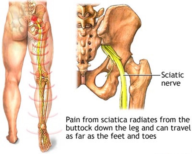
Sciatica refers to pain, weakness, numbness, or tingling in the leg. It is caused by injury to or pressure on the sciatic nerve. Sciatica is a symptom of another medical problem. It is not a medical condition on its own.
Sciatica occurs when there is pressure or damage to the sciatic nerve. This nerve starts in the lower back and runs down the back of each leg. This nerve controls the muscles of the back of the knee and lower leg. It also provides sensation to the back of the thigh, part of the lower leg, and the sole of the foot.
Sciatica pain can vary widely. It may feel like a mild tingling, dull ache, or burning sensation. In some cases, the pain is severe enough to make a person unable to move.
The pain most often occurs on one side. Some people have sharp pain in one part of the leg or hip and numbness in other parts. The pain or numbness may also be felt on the back of the calf or on the sole of the foot. The affected leg may feel weak. Sometimes, your foot gets caught on the ground when walking.
The pain often starts slowly. It may get worse:
· After standing or sitting
· At night
· When sneezing, coughing, or laughing
· When bending backward or walking more than a few yards, especially if caused by spinal stenosis
Because sciatica is a symptom of another medical condition, the underlying cause should be identified and treated.
In some cases, no treatment is required and recovery occurs on its own.
Conservative (non-surgical) treatment is best in many cases. Your doctor may recommend the following steps to calm your symptoms and reduce inflammation:
· Apply heat or ice to the painful area. Try ice for the first 48 to 72 hours, then use heat.
· Take over-the-counter pain relievers such as ibuprofen (Advil, Motrin IB) or acetaminophen (Tylenol).
· Reduce your activity for the first couple of days. Then, slowly start your usual activities.
· Do not do heavy lifting or twisting of your back for the first 6 weeks after the pain begins.
· Start exercising again after 2 to 3 weeks. Include exercises to strengthen your abdomen and improve flexibility of your spine.
Physical therapy may also be recommended. Additional treatments depend on the condition that is causing the sciatica.
If these measures do not help, your doctor may recommend injections of certain medicines to reduce swelling around the nerve. Other medicines may be prescribed to help reduce the stabbing pains due to nerve irritation.
Nerve pain is very difficult to treat. If you have ongoing problems with pain, you may want to see a neurologist or a pain specialist to ensure that you have access to the widest range of treatment options.
More serious complications depend on the cause of sciatica, such as slipped disc or spinal stenosis. Sciatica can lead to permanent numbness or weakness of your leg.
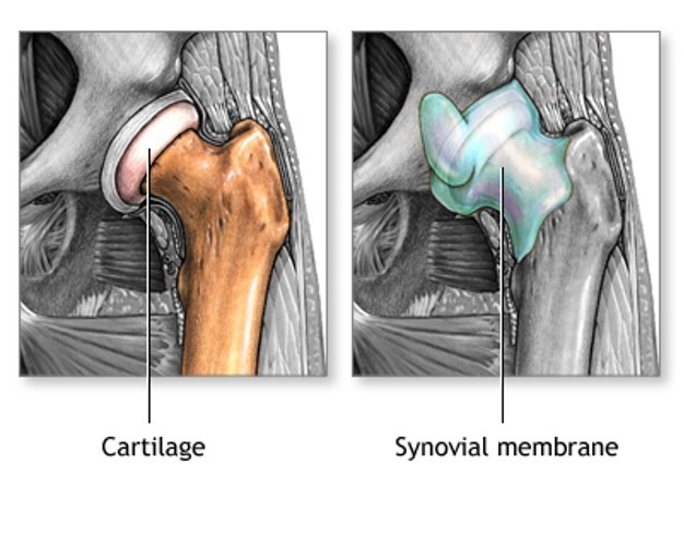
Arthritis involves the breakdown of cartilage. Normal cartilage protects a joint and allows it to move smoothly. Cartilage also absorbs shock when pressure is placed on the joint, such as when you walk. Without the normal amount of cartilage, the bones rub together. This causes swelling (inflammation), and stiffness.
Joint inflammation and damage may result from:
An autoimmune disease (the body's immune system mistakenly attacks healthy tissue)
Broken bone
General "wear and tear" on joints
Infection, most often by bacteria or virus
Crystals such as uric acid or calcium pyrophosphate dihydrate
In most cases, the joint inflammation goes away after the cause goes away or is treated. Sometimes, it does not. When this happens, you have long-term (chronic) arthritis.
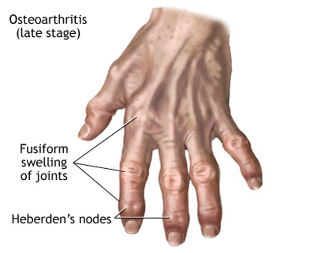
Arthritis may occur in men or women. Osteoarthritis is the most common type.
Other, more common types of inflammatory arthritis include:
The goal of treatment is to reduce pain, improve function, and prevent further joint damage. The underlying cause often cannot be cured.
LIFESTYLE CHANGES
Lifestyle changes are the preferred treatment for osteoarthritis and other types of joint swelling. Exercise can help relieve stiffness, reduce pain and fatigue, and improve muscle and bone strength. Your health care team can help you design an exercise program that is best for you.
Exercise programs may include:
Low-impact aerobic activity (also called endurance exercise). Walking is a good example.
Range of motion exercises for flexibility.
Strength training for muscle tone.
Your provider may suggest physical therapy. This might include:
Heat or ice
Splints or orthotics to support joints and help improve their position; this is often needed for rheumatoid arthritis
Water therapy
Massage
Other things you can do include:
Get plenty of sleep. Sleeping 8 to 10 hours a night and taking naps during the day can help you recover from a flare-up more quickly, and may even help prevent flare-ups.
Avoid staying in one position for too long.
Avoid positions or movements that place extra stress on your sore joints.
Change your home to make activities easier. For example, install grab bars in the shower, the tub, and near the toilet.
Try stress-reducing activities, such as meditation, yoga, or tai chi.
Eat a healthy diet full of fruits and vegetables, which contain important vitamins and minerals, especially vitamin E.
Eat foods rich in omega-3 fatty acids, such as cold water fish (salmon, mackerel, and herring), flaxseed, rapeseed (canola) oil, soybeans, soybean oil, pumpkin seeds, and walnuts.
Avoid excessive alcohol and smoking.
Apply capsaicin cream over your painful joints. You may feel improvement after applying the cream for 3 to 7 days.
Lose weight, if you are overweight. Weight loss can greatly improve joint pain in the legs and feet.
MEDICINES
Medicines may be prescribed along with lifestyle changes. All medicines have some risks. You should be closely followed by a doctor when taking arthritis medicines.
Over-the-counter medicines:
Acetaminophen (Tylenol) is often the first medicine tried. Take up to 3,000 mg a day (2 arthritis-strength Tylenol every 8 hours). To prevent damage to your liver, do not take more than the recommended dose. Since multiple medicines are available without a prescription that also contain acetaminophen, you will need to include them in the 3,000 mg per day maximum. Also, avoid alcohol when taking acetaminophen.
Aspirin, ibuprofen, or naproxen are nonsteroidal anti-inflammatory drugs (NSAIDs) that can relieve arthritis pain. However, they can carry risks when used for a long time. Possible side effects include heart attack, stroke, stomach ulcers, bleeding from the digestive tract, and kidney damage.
Prescription medicines:
Corticosteroids ("steroids") help reduce inflammation. They may be injected into painful joints or given by mouth.
Disease-modifying anti-rheumatic drugs (DMARDs) are used to treat autoimmune arthritis. They include methotrexate, sulfasalazine, hydroxychloroquine, and leflunomide.
Biologics are used for the treatment of autoimmune arthritis especially rheumatoid arthritis (RA). They include etanercept (Enbrel), infliximab (Remicade), adalimumab (Humira), abatacept (Orencia), rituximab (Rituxan), golimumab (Simponi), certolizumab (Cimzia), and tocilizumab (Actemra). These drugs can improve the quality of life for many people, but can have serious side effects.
Other drugs for RA -- Janus kinase inhibitor: Tofacitinib (Xeljanz). This is a medicine taken by mouth that is now approved for treating RA.
For gout, allopurinol (Zyloprim), febuxostat (Uloric) or probenecid (Benemid) may be used to lower uric acid.
It is very important to take your medicines as directed by your provider. If you are having problems doing so (for example, because of side effects), you should talk to your provider. Also make sure your provider knows about your all the medicines you are taking, including vitamins and supplements bought without a prescription.
SURGERY AND OTHER TREATMENTS
In some cases, surgery may be done if other treatments have not worked. This may include:
Early diagnosis and treatment can help prevent joint damage. If you have a family history of arthritis, tell your provider , even if you do not have joint pain.
Avoiding excessive, repeated motions may help protect you against osteoarthritis.