.

кожа | zdrav.kz
X

Электрондық поштаңызға соңғы жаңалықтарды алыңыз

X

Получайте самые последние новости на свой e-mail

кожа

Жалпы ақпарат

Себептері

Симптомдары

Диагностика

Емдеу

Болжамы

Мүмкін асқынулары

Қашан дәрігерге көріну қажет

Алдын алу

Синонимдары


Жалпы ақпарат

Себореялық дерматит – терінің жиі кездесетін, қабыну ауруы. Бұл кезде шаштың түбіндегі, бет пен құлақтың май безі жақсы дамыған бөліктерінде ақ-сары тұстары болады. олар өзгермеген немесе қызармаған тұста пайда болуы мүмкін.

«Бесік телпегі» - сәбилерде себореялық дерматит кезіндегі бастың терісінің зақымдануын сипаттайтын термин.


Себептері

Себореялық дерматиттің нақты себебі белгісіз. Дәрігерлердің ойынша, ол бірнеше себептің бірігуінен болуы мүмкін, гормоналдық үйлесімнің бұзылуын, иммундық жүйенің әлсіреуін, қоректік заттектердің жеткіліксіздігін, немесе жүйке жүйесінің ауруларын қоса.

Malassezia зеңдерінен пайда болатын тітіркеніс те осы жағдайға әкелуі мүмкін. Себореялық дерматит тұқым қуалауы мүмкін.


Терінің қабаттары

Қауіп факторлары:

  • Күйзеліс немесе шаршау
  • Экстремалдық ауа райы
  • Майлы тері немесе терідегі безеулер
  • Кейбір сусабындар немесе теріге күтім жасау құралы
  • Спирт құрамдас лосьондар қолдану
  • Семіздік
  • Неврологиялық аурулар, оның ішінде, бассүйек-ми жарақаты, Паркинсон ауруы немесе инсульт
  • Адамның иммунитет тапшылығы вирусы (ВИЧ)


Симптомдары

Себореялық дерматит дененің әр бөлігінде пайда болуы мүмкін. Әдетте, ол терінің май безі жақсы дамыған тұстарында пайда болады. Көбінесе, бұл бастың, қабақтың терісі, мұрын-ерін қыртысы, құлақ артындағы, сыртқы құлақтағы тұстар, сондай-ақ, кеуде қуысы.

Себореялық дерматиттің симптомдары:

•      Терінің зақымдануы

•      Терінің үлкен аумағындағы түйіндақтар

•      Терінің майлы бөліктері

•      Теріде ақ, қабыршақтанған, сары, майлы бөліктердің пайда болуы

•      Жұқпаланған кезде нашарлауы мүмкін қышу

•      Терінің аздаған қызаруы

•      Шаштың түсуі


Диагностика

Диагноз тері зақымдануының өзіне тән сыртқы түріне және орналасуына сай қойылады. Әрі қарай, тері биопсиясы секілді тексерулер сирек қажет болады.


Емдеу

Қабыршақтану мен құрғақтықты қайызғаққа арналған арнайы сусабынның көмегімен емдеуге болады. оларды дәріханада дәрігердің рецептінісіз сатып алуға болады. жапсырмасында себореялық дерматитті емдейді деп жазылған сусабынды іздеңіз. Олардың құрамында салицил қышқылы, таскөмір шайыры, мырыш, резорцин, кетоконазол немесе селен сульфиді секілді құрамбөліктер болады. сусабындарды нұсқаулығында көрсетілуіне сәйкес қолданыңыз.  

Аурудың ауыр өтуінде дәрігер селен сульфидінің, кетоконазол немесе кортикостероидтардың мөлшері жоғары сусабынды немесе лосьонды қолдануды тағайындауы мүмкін.  Сондай-ақ, құрамында иммунитетті ынталандырғыш болатын крем ұсынылуы мүмкін. Бұл дәрі иммундық жүйенің жұмысын басуға қабілетті, осылайша, қабынуды азайтады.

Күн сәулесінің әсер етуінде себореялық дерматиттің барысы жақсарады деп саналады. Кейбір адамдар жазда жағдай жақсаратындығын айтады, әсіресе, белсенді демалыстан кейін.  


Болжамы

Себореялық дерматит – созылмалы ауру (өмір бойғы), асқыну мен басылу кзеңдері алмасады, бірақ, оның симптомдарын сәйкес емдеумен бақылауға болады.

Себореялық дерматиттегі жағдайдың ауырлығы қауіп факторларының әсерін жоюда және теріге күтім жасау қағидаларын ұстануда жеңілдейді. 


Мүмкін асқынулары

·         Психологиялық күйзеліс, өзін өзі бағалаудың төмендеуі, ұялып-қысылу

·         Екінші бактериалдық немесе зеңдік инфекцияның қосылуы.


Қашан дәрігерге көріну қажет

Дәрігерден кеңес алыңыз, егер себореялық дерматиттің симптомдары үй жағдайында емдеуге және дәрігердің рецептінсіз босатылатын дәрі препараттарын қабылдауға серпін бермесе. 

Сондай-ақ, дәрігерге көрініңіз, егер зақымдану бөліктерінің бетінде сұйықтық, ірің немесе қабық пайда болса, немесе бұл тұстар қызарып, ауырсынатын болса.   


Алдын алу

Себореялық дерматит кезіндегі жағдайдың ауырлығы қауіп факторларының әсерін жоюда және теріге күтім жасау қағидаларын ұстануда жеңілдейді.


Синонимдары

Қайызғақ; Себореялық экзема; «Бесік телпегі».


Ақпарат көзі: АҚШ Ұлттық денсаулық институттарының мәліметтер қоры:http://www.nlm.nih.gov/medlineplus/ency/article/000963.htm

Аударған:АсельСтамбекова, HealthСity жобасының дербес үйлестірушісі

Редакциялық алқа:

Алмаз Шарман, медицина профессоры 

Солнцезащитные крема предотвращают старение кожи

Согласно проведенному в Австралии исследованию, обычный крем от загара способен предотвратить процесс старения кожи, которое происходит из-за воздействия ультрафиолетовых лучей (солнечной геродермии).

Contents

Considerations

Causes

Symptoms

First Aid

DO NOT

When to Contact a Medical Professional

Prevention


Burns commonly occur by direct or indirect contact with heat, electric current, radiation, or chemical agents. Burns can lead to cell death.

Considerations

There are 3 levels of burns:

·         First-degree burns affect only the outer layer of the skin. They cause pain, redness, and swelling.

·         Second-degree burns affect both the outer and underlying layer of skin. They cause pain, redness, swelling, and blistering. They are also called partial thickness burns.

·         Third-degree burns affect the deep layers of skin. They are also called full thickness burns. They cause white or blackened, burned skin. The skin may be numb.

Burns fall into 2 groups.

Minor burns are:

·         First degree burns anywhere on the body

·         Second degree burns less than 2 to 3 inches (5 to 7.5 centimeters) wide 

Major burns include:

·         Third-degree burns

·         Second-degree burns more than 2 to 3 inches (5 to 7.5 centimeters) wide

·         Second-degree burns on the hands, feet, face, groin, buttocks, or over a major joint

You can have more than 1 type of burn at a time.

Severe burns need urgent medical care. This can help prevent scarring, disability, and deformity.

Burns on the face, hands, feet, and genitals can be particularly serious.

Children under age 4 and adults over age 60 have a higher chance of complications and death from severe burns because their skin tends to be thinner than in other age groups.

Causes

Causes of burns from most to least common are:

·         Fire/flame

·         Scalding from steam or hot liquids

·         Touching hot objects

·         Electrical burns

·         Chemical burns

Burns can be the result of any of the following:

·         House and industrial fires

·         Car accidents

·         Playing with matches

·         Faulty space heaters, furnaces, or industrial equipment

·         Unsafe use of firecrackers and other fireworks

·         Kitchen accidents, such as a child grabbing a hot iron or touching the stove

You can also burn your airways if you breathe in smoke, steam, superheated air, or chemical fumes in poorly ventilated areas.

Symptoms

Burn symptoms can include:

·         Blisters that are either intact (unbroken) or have ruptured and are leaking fluid

·         Pain (How much pain you have is unrelated to the level of burn. The most serious burns can be painless.)

·         Peeling skin

·         Shock (Watch for pale and clammy skin, weakness, blue lips and fingernails, and a drop in alertness.)

·         Swelling

·         Red, white, or charred skin

If you have burned your airways, you may have:

·         Burns on the head, face, neck, eyebrows, or nose hairs

·         Burned lips and mouth

·         Coughing

·         Difficulty breathing

·         Dark, black-stained mucus

·         Voice changes

·         Wheezing

First Aid

Before giving first aid, it is important to determine what type of burn the person has. If you are not sure, treat it as a major burn. Serious burns need medical care right away. Call your local emergency number or 911.

MINOR BURNS

If the skin is unbroken:

·         Run cool water over the area of the burn or soak it in a cool water bath (not ice water). Keep the area under water for at least 5 minutes. A clean, cold, wet towel will help reduce pain.

·         Calm and reassure the person.

·         After flushing or soaking the burn, cover it with a dry, sterile bandage or clean dressing.

·         Protect the burn from pressure and friction.

·         Over-the-counter ibuprofen or acetaminophen can help relieve pain and swelling. Do NOT give aspirin to children under 12.

·         Once the skin has cooled, moisturizing lotion also can help.

Minor burns will often heal without further treatment. Make sure the person is up to date on their tetanus immunization.

MAJOR BURNS

If someone is on fire, tell the person to stop, drop, and roll. Then, follow these steps:

·         Wrap the person in thick material; such as a wool or cotton coat, rug, or blanket. This helps put out the flames.

·         Pour water on the person.

·         Call 911 or your local emergency number.

·         Make sure that the person is no longer touching any burning or smoking materials.

·         Do NOT remove burned clothing that is stuck to the skin.

·         Make sure the person is breathing. If necessary, begin rescue breathing and CPR.

·         Cover the burn area with a dry sterile bandage (if available) or clean cloth. A sheet will do if the burned area is large. Do NOT apply any ointments. Avoid breaking burn blisters.

·         If fingers or toes have been burned, separate them with dry, sterile, non-sticky bandage.

·         Raise the body part that is burned above the level of the heart.

·         Protect the burn area from pressure and friction.

·         If an electrical injury may have caused the burn, DO NOT touch the victim directly. Use a non-metallic object to move the person away from exposed wires before starting first aid.

You will also need to prevent shock. If the person does not have a head, neck, back, or leg injury, follow these steps:

·         Lay the person flat

·         Raise the feet about 12 inches (30 centimeters)

·         Cover the person with a coat or blanket

Continue to monitor the person's pulse, rate of breathing, and blood pressure until medical help arrives.

DO NOT

Things that should not be done for burns include:

·         Do NOT apply ointment, butter, ice, medicines, cream, oil spray, or any household remedy to a severe burn.

·         Do NOT breathe, blow, or cough on the burn.

·         Do NOT disturb blistered or dead skin.

·         Do NOT remove clothing that is stuck to the skin.

·         Do NOT give the person anything by mouth, if there is a severe burn.

·         Do NOT place a severe burn in cold water. This can cause shock.

·         Do NOT place a pillow under the person's head if there is an airways burn. This can close the airways.

When to Contact a Medical Professional

Call 911 or your local emergency number if:

·         The burn is very large, about the size of your palm or larger.

·         The burn is severe (third degree).

·         You aren't sure how serious it is.

·         The burn is caused by chemicals or electricity.

·         The person shows signs of shock.

·         The person breathed in smoke.

·         Physical abuse is the known or suspected cause of the burn.

·         There are other symptoms associated with the burn.

For minor burns, call your health care provider if you still have pain after 48 hours.

Call a provider right away if signs of infection develop. These signs include:

·         Drainage or pus from the burned skin

·         Fever

·         Increased pain

·         Red streaks spreading from the burn

·         Swollen lymph nodes

Also call a provider right away if symptoms of dehydration occur with a burn:

·         Decreased urination

·         Dizziness

·         Dry skin

·         Headache

·         Lightheadedness

·         Nausea (with or without vomiting)

·         Thirst

Children, older people, and anyone with a weakened immune system (for example, from HIV) should be seen right away.

The provider will perform a history and physical examination. Tests and procedures will be done as needed.

These may include:

·         Airway and breathing support, including a face mask, tube through the mouth into the trachea, or breathing machine (ventilator) for serious burns or those involving the face or airway

·         Blood and urine tests if shock or other complications are present

·         Chest x-ray for face or airway burns

·         EKG (electrocardiogram, or heart tracing), if shock or other complications are present

·         Intravenous fluids (fluids through a vein), if shock or other complications are present

·         Medicines for pain relief and to prevent infection

·         Ointments or creams applied to the burned areas

·         Tetanus immunization, if not up to date

The outcome will depend on the type (degree), extent, and location of the burn. It also depends upon whether internal organs have been affected, and if other trauma has occurred. Burns can leave permanent scars. They can also be more sensitive to temperature and light than normal skin. Sensitive areas, such as the eyes, nose, or ears, may be badly injured and have lost normal function.

With airway burns, the person may have less breathing capacity and permanent lung damage. Severe burns that affect the joints may result in contractures, leaving the joint with decreased movement and a reduction in function.

Prevention

To help prevent burns:

·         Install smoke alarms in your home. Check and change batteries regularly.

·         Teach children about fire safety and the danger of matches and fireworks.

·         Keep children from climbing on top of a stove or grabbing hot items such as irons and oven doors.

·         Turn pot handles toward the back of the stove so that children can't grab them and they can't accidentally be knocked over.

·         Place fire extinguishers in key locations at home, work, and school.

·         Remove electrical cords from floors and keep them out of reach.

·         Know about and practice fire escape routes at home, work, and school.

·         Set the water heater temperature at 120°F (48.8°C) or less.


Sourcehttps://medlineplus.gov/ency/article/000030.htm

The breast is made up of glands called lobules that can make milk and thin tubes called ducts that carry the milk from the lobules to the nipple. Breast tissue also contains fat and connective tissue, lymph nodes, and blood vessels.

Inside a woman’s breast are 15 to 20 sections (lobes). Each lobe is made of many smaller sections (lobules). Lobules have groups of tiny glands that can make milk. After a baby is born, breast milk flows from the lobules through thin tubes (ducts) to the nipple. Fibrous tissue and fat fill the spaces between the lobules and ducts.

Your skin is your body's largest organ. It covers and protects your body. Your skin

·         Holds body fluids in, preventing dehydration

·         Keeps harmful microbes out, preventing infections

·         Helps you feel things like heat, cold, and pain

·         Keeps your body temperature even

·         Makes vitamin D when the sun shines on it

Anything that irritates, clogs, or inflames your skin can cause symptoms such as redness, swelling, burning, and itching. Allergies, irritants, your genetic makeup, and certain diseases and immune system problems can cause rasheshives, and other skin conditions. Many skin problems, such as acne, also affect your appearance.

Contents

Causes

Symptoms

Exams and Tests

Treatment

Outlook (Prognosis)

Possible Complications

When to Contact a Medical Professional

Prevention


Newborn jaundice is when a baby has high levels of bilirubin in the blood. Bilirubin is a yellow substance that the body creates when it replaces old red blood cells. The liver helps break down the substance so it can be removed from the body in the stool.

High levels of bilirubin makes your baby's skin and whites of the eyes look yellow. This is called jaundice.

Newborn jaundice (producing yellow skin) can have many causes, but the majority of these infants have a condition called physiological jaundice, a natural occurrence in the newborn due to the immature liver. This type of jaundice is short term, generally lasting only a few days. Jaundice should be evaluated by a physician until decreasing or normal levels of bilirubin are measured in the blood.

Causes

 It is normal for a baby's bilirubin level to be a bit higher after birth.

When the baby is growing in the mother's womb, the placenta removes bilirubin from the baby's body. The placenta is the organ that grows during pregnancy to feed the baby. After birth, the baby's liver starts doing this job. This can take a while.

Most newborns have some yellowing of the skin, or jaundice. This is called  "physiological jaundice." It is harmless, and usually is worst when the baby is 2 - 4 days old. It goes away within 2 weeks and doesn't usually cause a problem.  

Two types of jaundice may occur in newborns who are breast fed. Both types are usually harmless.

  • Breastfeeding jaundice is seen in breastfed babies during the first week of life, especially in babies who do not nurse well or if the mother's milk is slow to come in.
  • Breast milk jaundice may appear in some healthy, breastfed babies after day 7 of life. It usually peaks during weeks 2 and 3. It may last at low levels for a month or more. It may be due to how substances in the breast milk affect how bilirubin breaks down in the liver. Breast milk jaundice is different than breastfeeding jaundice.

Severe newborn jaundice may occur if your baby has a condition that increases the number of red blood cells that need to be replaced in the body, such as:

  • Abnormal blood cell shapes
  • Blood type mismatch between the mother and the baby
  • Bleeding underneath the scalp (cephalohematoma) caused by a difficult delivery
  • Higher levels of red blood cells, which is more common in small-for-gestational-age babies and some twins
  • Infection
  • Lack (deficiency) of certain important proteins, called enzymes

Things that make it harder for the baby's body to remove bilirubin may also lead to more severe jaundice, including:

  • Certain medications
  • Congenital infections, such as rubella, syphilis, and others
  • Diseases that affect the liver or biliary tract, such as cystic fibrosis or hepatitis
  • Low oxygen level (hypoxia)
  • Infections (such as sepsis)
  • Many different genetic or inherited disorders

Babies who are born too early (premature) are more likely to develop jaundice than full-term babies.

Symptoms

Jaundice causes a yellow color of the skin. The color sometimes begins on the face and then moves down to the chest, belly area, legs, and soles of the feet.

Sometimes, infants with significant jaundice have extreme tiredness and poor feeding.

Exams and Tests

Doctors, nurses, and family members will watch for signs of jaundice at the hospital, and after the newborn goes home.

Any infant who appears jaundiced should have bilirubin levels measured right away. This can be done with a blood test.

Many hospitals check total bilirubin levels on all babies at about 24 hours of age. Hospitals use probes that can estimate the bilirubin level just by touching the skin. High readings need to be confirmed with blood tests.

Tests that will likely be done include:

  • Complete blood count
  • Coomb's test
  • Reticulocyte count

Further testing may be needed for babies who need treatment or whose total bilirubin levels are rising more quickly than expected.

Treatment

Treatment is usually not needed.

When determining treatment, the doctor must consider:

  • The baby's bilirubin level
  • How fast the level has been rising
  • Whether the baby was born early (babies born early are more likely to be treated at lower bilirubin levels)
  • How old the baby is now

Your child will need treatment if the bilirubin level is too high or is rising too quickly.

Keep the baby well hydrated with breast milk or formula. Frequent feedings (up to 12 times a day) encourage frequent bowel movements, which help remove bilirubin through the stools. Ask your doctor before giving your newborn extra formula.

Some newborns need to be treated before they leave the hospital. Others may need to go back to the hospital when they are a few days old. Treatment in the hospital usually lasts 1 to 2 days.

Sometimes special blue lights are used on infants whose levels are very high. This is called phototherapy. These lights work by helping to break down bilirubin in the skin.

The infant is placed under artificial light in a warm, enclosed bed to maintain constant temperature. The baby will wear only a diaper and special eye shades to protect the eyes. The American Academy of Pediatrics recommends that breastfeeding be continued through phototherapy, if possible. Rarely, the baby may have an intravenous (IV) line to deliver fluids.

If the bilirubin level is not too high or is not rising quickly, you can do phototherapy at home with a fiberoptic blanket, which has tiny bright lights in it. You may also use a bed that shines light up from the mattress.

  • You must keep the light therapy on your child's skin and feed your child every 2 to 3 hours (10 to 12 times a day).
  • A nurse will come to your home to teach you how to use the blanket or bed, and to check on your child.
  • The nurse will return daily to check your child's weight, feedings, skin, and bilirubin levels.
  • You will be asked to count the number of wet and dirty diapers.

In the most severe cases of jaundice, an exchange transfusion is required. In this procedure, the baby's blood is replaced with fresh blood. Treating severely jaundiced babies with intravenous immunoglobulin may also be very effective at reducing bilirubin levels.

Outlook (Prognosis)

Usually newborn jaundice is not harmful. For most babies, jaundice usually gets better without treatment within 1 to 2 weeks.

Very high levels of bilirubin can damage the brain. This is called kernicterus. However, the condition is almost always diagnosed before levels become high enough to cause this damage.

For babies who need treatment, the treatment is usually effective.

Possible Complications

Rare, but serious, complications from high bilirubin levels include:

  • Cerebral palsy
  • Deafness
  • Kernicterus -- brain damage from very high bilirubin levels

When to Contact a Medical Professional

All babies should be seen by a health care provider in the first 5 days of life to check for jaundice.

  • Those who spend less than 24 hours in a hospital should be seen by age 72 hours.
  • Infants sent home between 24 and 48 hours should be seen again by age 96 hours.
  • Infants sent home between 48 and 72 hours should be seen again by age 120 hours.

Jaundice is an emergency if the baby has a fever, has become listless, or is not feeding well. Jaundice may be dangerous in high-risk newborns.

Jaundice is generally NOT dangerous in term, otherwise healthy newborns. Call the infant's health care provider if:

  • Jaundice is severe (the skin is bright yellow)
  • Jaundice continues to increase after the newborn visit, lasts longer than 2 weeks, or other symptoms develop
  • The feet, especially the soles, are yellow

Prevention

In newborns, some degree of jaundice is normal and probably not preventable. The risk of significant jaundice can often be reduced by feeding babies at least 8 to 12 times a day for the first several days and by carefully identifying infants at highest risk.

All pregnant women should be tested for blood type and unusual antibodies. If the mother is Rh negative, follow-up testing on the infant's cord is recommended. This may also be done if the mother's blood type is O+, but it is not needed if careful monitoring takes place.

Careful monitoring of all babies during the first 5 days of life can prevent most complications of jaundice. Ideally, this includes:

  • Considering a baby's risk for jaundice
  • Checking bilirubin level in the first day or so
  • Scheduling at least one follow-up visit the first week of life for babies sent home from the hospital in 72 hours


Source: http://www.nlm.nih.gov/medlineplus/ency/article/001559.htm

Ученые приблизились к созданию лекарства от облысения

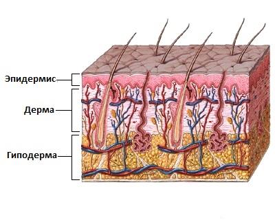
Команда исследователей из Колумбийского университета города Нью-Йорка впервые в истории медицины разработала способ возобновления роста волос.

Как уверяют ученые, благодаря открытию, в будущем медики смогут спровоцировать процесс роста собственных волос у любого человека.

Мазмұны

Жалпы ақпарат

Себептері

Симптомдары

Емдеу

Болжамы


Жалпы ақпарат

Эризипелоид – терінің бактериалдық қабынуы (дерматит).


Себептері

Эризипелоидтің қоздырғышы - Erysipelothrix Rhusiopathiae бактериясы.  Бактериялардың бұл түрі балықта, құста, сүт қоректілерде, моллюскілер мен шаян тәрізділерде мекендейді. Адамдар әдетте, осы жануарлармен жұмыс істегенде жұқтырады (мысалы, фермерлер немесе қасапшылар).


Симптомдары

Негізгі симптомы – терінің сезімталдығының артуы және қызаруы.


Емдеу

Ауруды емдеу үшін антибактериалдық препараттар тағайындалады, әсіресе, пенициллин.


Болжамы

Инфекция сирек таралады және өздігінше өтуі мүмкін. 

Солнечный свет улучшает здоровье и продлевает жизнь

Солнечный светСогласно исследованию, проведенном в Эдинбургском Университете, загар может снизить артериальное давление, уменьшить риск сердечного приступа и инсульта, а также продлить жизнь.

Squamous cell cancer is a common type of skin cancer.

Other common types of skin cancer are:

·         Basal cell

·         Melanoma


Causes

Squamous cell cancer may occur in undamaged skin. Or it can occur in skin that has been injured or inflamed. Most squamous cell carcinomas occur on skin that is regularly exposed to sunlight or other ultraviolet radiation.

The earliest form of squamous cell cancer is called Bowen disease (or squamous cell carcinoma in situ). This type does not spread to nearby tissues.

Actinic keratosis is a precancerous skin lesion that may become a squamous cell cancer.

Risks of squamous cell cancer include:

·         Having light-colored skin, blue or green eyes, or blond or red hair

·         Long-term, daily sun exposure (such as in people who work outside)

·         Many severe sunburns early in life

·         Older age

·         Having had many x-rays

·         Chemical exposure


Symptoms

Squamous cell cancer usually occurs on the face, ears, neck, hands, or arms. It may occur on other areas.

The main symptom is a growing bump that may have a rough, scaly surface and flat reddish patches.

The earliest form (squamous cell carcinoma in situ) can appear as a scaly, crusted, and large reddish patch that can be larger than 1 inch.

A sore that does not heal can be a sign of squamous cell cancer. Any change in an existing wart,mole, or other skin lesion could be a sign of skin cancer.


Exams and Tests

Your doctor will check your skin and look at the size, shape, color, and texture of any suspicious areas.

If your doctor thinks you might have skin cancer, a piece of skin will be removed. This is called a skin biopsy. The sample is sent to a lab for examination under a microscope.

A skin biopsy must be done to confirm squamous cell skin cancer or other skin cancers.


Treatment

Treatment depends on the size and location of the skin cancer, how far it has spread, and your overall health. Some squamous cell skin cancers may be more difficult to treat.

Treatment may involve:

·         Excision: Cutting out the skin cancer and stitching the skin together.

·         Curettage and electrodessication: Scraping away cancer cells and using electricity to kill any that remain; it is used to treat cancers that are not very large or deep.

·         Cryosurgery: Freezing the cancer cells, which kills them. This is used for small and superficial cancers.

·         Medication: Skin creams containing imiquimod or 5-fluorouracil for superficial (not very deep) squamous cell cancer.

·         Mohs surgery: Removing a layer of skin and looking at it immediately under a microscope, then removing layers of skin until there are no signs of the cancer; usually used for skin cancers on the nose, ears, and other areas of the face.

·         Photodynamic therapy: Treatment using light may be used to treat superficial cancers.

·         Radiation may be used if squamous cell cancer has spread to organs or lymph nodes or if the cancer cannot be treated with surgery.


Outlook (Prognosis)

How well a person does depends on many things, including how soon the cancer was diagnosed. Most of these cancers are cured when treated early.

Some squamous cell cancers may return.


Possible Complications

Squamous cell cancer spreads faster than basal cell cancer, but still may grow slowly. It may spread to other parts of the body, including internal organs.


When to Contact a Medical Professional

Call for an appointment with your health care provider if you have a sore or spot on your skin that changes in:

·         Appearance

·         Color

·         Size

·         Texture

Also call if a spot becomes painful or swollen or if it starts to bleed or itch.


Prevention

The American Cancer Society recommends that a health care provider examine your skin every year if you are older than 40 and every 3 years if you are 20 to 40 years old. You should also examine your own skin once a month.

If you have had skin cancer, you should have regular checkups so that a doctor can examine your skin. You should also check your own skin once a month. Use a hand mirror for hard-to-see places. Call your doctor if you notice anything unusual.

The best way to prevent skin cancer is to reduce your exposure to sunlight. Always use sunscreen:

·         Apply sunscreen with sun protection factor (SPF) of at least 30, even when you are going outdoors for a short time.

·         Apply a large amount of sunscreen on all exposed areas, including ears and feet.

·         Look for sunscreen that blocks both UVA and UVB light.

·         Use a water-resistant sunscreen.

·         Apply sunscreen at least 30 minutes before going out. Follow package instructions about how often to reapply. Be sure to reapply after swimming or sweating.

·         Use sunscreen in winter and on cloudy days, too.

Other measures to help you avoid too much sun exposure:

·         Ultraviolet light is most intense between 10 a.m. and 4 p.m. So try to avoid the sun during these hours.

·         Protect the skin by wearing wide-brim hats, long-sleeve shirts, long skirts, or pants.

·         Avoid surfaces that reflect light more, such as water, sand, concrete, and areas that are painted white.

·         The higher the altitude, the faster your skin burns.

·         Do not use sun lamps and tanning beds (salons). Spending 15 to 20 minutes at a tanning salon is as dangerous as a day spent in the sun.


Alternative Names

Cancer - skin - squamous cell; Skin cancer - squamous cell; Nonmelanoma skin cancer - squamous cell; NMSC - squamous cell; Squamous cell skin cancer; Squamous cell carcinoma of the skin


https://www.nlm.nih.gov/medlineplus/ency/article/000829.htm