Көзге түсетін бөгде заттардың көпшілігі (нысандар) – топырақ, кірпік немесе киімнің қылдары секілді шағын бөлшектер. Қауіптісі – металдың бөлшектері немесе басқа нысандар, көзге үлкен жылдамдықпен түсетін. Олар механикалық аспаптардан, газоншапқыштан немесе қарудан ұшқан заттар болуы мүмкін. Олар күрделі жарақат туындатуы мүмкін.
Көздің жарақаты қан кетуін, қарашықтың өлшемінің өзгеруін, көзбұршақтың бұлыңғырлануын, көрудің нашарлауын немесе көрудің өзгеруін туындатуы мүмкін. Бөгде заттар көзге терең енуі мүмкін. Мұндайда оларды қозғамау қажет. Оларды көз маманы алуы тиіс (офтальмолог).
Шағын өлшемді нысандарды алып тастау үшін не істеу қажет? Алдымен, көзді сүртуге болмайды. Қолды мұқият жуу қажет. Жақсы жарықта көзді мұқият қарау қажет. Егер нысан көзде көрінсе, оны сумен шаюға болады. нысан жоғарғы немесе төменгі қабақта болуы мүмкін. Мұндайда қабақты ашып тұрып, мақта тығынды қолданумен қабақ астынан затты абайлап алып тастауға болады. көздің үстінен заттарды алып тастауға мақта тығынды қолдануға болмайды.
Егер бөгде затты көзден үй жағдайында алып тастауға келмесе, көзді таза таңғышпен жауып (көзді қыспаңыз), дәрігерге көріну қажет. Егер бөгде затты алып тастасаңыз, бірақ, біраздан кейін көзде бірнәрсе қалғандай сезім пайда болса, мөлдірқабық зақымдануы мүмкін. Бұл жағдайда дәрігерге міндетті түрде бару қажет.
Көзге енген заттарды алып тастау үшін не істеу қажет? Затты көзде қалдыру қажет, оны ҚОЗҒАУҒА БОЛМАЙДЫ. Көзді таза таңғышпен жауып (көзді қыспаңыз), шұғыл түрде дәрігерге көріну қажет.
If more pressure is put on a bone than it can stand, it will split or break. A break of any size is called a fracture. If the broken bone punctures the skin, it is called an open fracture (compound fracture).
A stress fracture is a hairline crack in the bone that develops because of repeated or prolonged forces against the bone.
1. Check the person's airway and breathing. If necessary, call 911 and begin rescue breathing, CPR, or bleeding control.
2. Keep the person still and calm.
3. Examine the person closely for other injuries.
4. In most cases, if medical help responds quickly, allow the medical personnel to take further action.
5. If the skin is broken, it should be treated immediately to prevent infection. Call emergency help right away. DO NOT breathe on the wound or probe it. If possible, lightly rinse the wound to remove visible dirt or other contamination, but do not vigorously scrub or flush the wound. Cover with sterile dressings.
6. If needed, immobilize the broken bone with a splint or sling. Possible splints include a rolled up newspaper or strips of wood. Immobilize the area both above and below the injured bone.
7. Apply ice packs to reduce pain and swelling.
8. Take steps to prevent shock. Lay the person flat, elevate the feet about 12 inches (30 centimeters) above the head, and cover the person with a coat or blanket. However, DO NOT move the person if a head, neck, or back injury is suspected.
CHECK BLOOD CIRCULATION
Check the person's blood circulation. Press firmly over the skin beyond the fracture site. (For example, if the fracture is in the leg, press on the foot). It should first blanch white and then "pink up" in about two seconds. Signs that circulation is inadequate include pale or blue skin, numbness or tingling, and loss of pulse.
If circulation is poor and trained personnel are NOT quickly available, try to realign the limb into a normal resting position. This will reduce swelling, pain, and damage to the tissues from lack of blood.
TREAT BLEEDING
Place a dry, clean cloth over the wound to dress it.
If the bleeding continues, apply direct pressure to the site of bleeding. DO NOT apply a tourniquet to the extremity to stop the bleeding unless it is life-threatening.
· DO NOT move the person unless the broken bone is stable.
· DO NOT move a person with an injured hip, pelvis, or upper leg unless it is absolutely necessary. If you must move the person, pull the person to safety by his clothes (such as by the shoulders of a shirt, a belt, or pant-legs).
· DO NOT move a person who has a possible spine injury.
· DO NOT attempt to straighten a bone or change its position unless blood circulation appears hampered.
· DO NOT try to reposition a suspected spine injury.
· The person is not responding or is losing consciousness.
· There is a suspected broken bone in the head, neck, or back.
· There is a suspected broken bone in the hip, pelvis, or upper leg.
· You cannot completely immobilize the injury at the scene by yourself.
· There is severe bleeding.
· An area below the injured joint is pale, cold, clammy, or blue.
· There is a bone projecting through the skin.
Even though other broken bones may not be medical emergencies, they still deserve medical attention. Call your health care provider to find out where and when to be seen.
If a young child refuses to put weight on an arm or leg after an accident, won't move the arm or leg, or you can clearly see a deformity, assume the child has a broken bone and get medical help.
Take the following steps to reduce your risk of a broken bone:
· Wear protective gear while skiing, biking, roller blading, and participating in contact sports. This includes using a helmet, elbow pads, knee pads, and shin pads.
· Create a safe home for young children. Place a gate at stairways and keep windows closed.
· Teach children how to be safe and look out for themselves.
· Supervise children carefully. There is no substitute for supervision, no matter how safe the environment or situation appears to be.
· Prevent falls by not standing on chairs, counter tops, or other unstable objects. Remove throw rugs and electrical cords from floor surfaces. Use handrails on staircases and non-skid mats in bathtubs. These steps are especially important for the elderly.
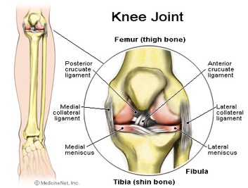
The knee is a joint which has three parts. The thigh bone (femur) meets the large shin bone (tibia) forming the main knee joint. This joint has an inner (medial) and an outer (lateral) compartment. The kneecap (patella) joins the femur to form a third joint, called the patellofemoral joint.
The knee joint is surrounded by a joint capsule with ligaments strapping the inside and outside of the joint (collateral ligaments) as well as crossing within the joint (cruciate ligaments). These ligaments provide stability and strength to the knee joint.
The meniscus is a thickened cartilage pad between the two joints formed by the femur and tibia. The meniscus acts as a smooth surface for the joint to move on. The knee joint is surrounded by fluid-filled sacs called bursae, which serve as gliding surfaces that reduce friction of the tendons. There is a large tendon (patellar tendon) which envelopes the knee cap and attaches to the front of the tibia bone. There are large blood vessels passing through the area behind the knee (referred to as the popliteal space). The large muscles of the thigh move the knee. In the front of the thigh, the quadriceps muscles extend, or straighten, the knee joint by pulling on the patellar tendon. In the back of the thigh, the hamstring muscles flex, or bend, the knee. The knee also rotates slightly under guidance of specific muscles of the thigh.
The knee functions to allow movement of the leg and is critical to normal walking. The knee flexes normally to a maximum of 135 degrees and extends to 0 degrees. The bursae, or fluid-filled sacs, serve as gliding surfaces for the tendons to reduce the force of friction as these tendons move. The knee is a weight-bearing joint. Each meniscus serves to evenly load the surface during weight-bearing and also aids in disbursing joint fluid for joint lubrication.
Injury can affect any of the ligaments, bursae, or tendons surrounding the knee joint. Injury can also affect the ligaments, cartilage, menisci (plural for meniscus), and bones forming the joint. The complexity of the design of the knee joint and the fact that it is an active weight-bearing joint are factors in making the knee one of the most commonly injured joints.
Trauma can cause injury to the ligaments on the inner portion of the knee (medial collateral ligament), the outer portion of the knee (lateral collateral ligament), or within the knee (cruciate ligaments). Injuries to these areas are noticed as immediate pain, but are sometimes difficult to localize. Usually, a collateral ligament injury is felt on the inner or outer portions of the knee. A collateral ligament injury is often associated with local tenderness over the area of the ligament involved. A cruciate ligament injury is felt deep within the knee. It is sometimes noticed with a "popping" sensation with the initial trauma. A ligament injury to the knee is usually painful at rest and may be swollen and warm. The pain is usually worsened by bending the knee, putting weight on the knee, or walking. The severity of the injury can vary from mild (minor stretching or tearing of the ligament fibers, such as a low grade sprain) to severe (complete tear of the ligament fibers). Patients can have more than one area injured in a single traumatic event.
Ligament injuries are initially treated with ice packs and immobilization, with rest and elevation. At first, it is generally recommended to avoid bearing weight on the injured joint and crutches may be required for walking. Some patients are placed in splints or braces to immobilize the joint to decrease pain and promote healing. Arthroscopic or open surgery may be necessary to repair severe injuries.
Surgical repair of ligaments can involve suturing, grafting, and synthetic graft repair. These procedures can be done by either open knee surgery or arthroscopic surgery (described in the section below). The decision to perform various types of surgery depends on the level of damage to the ligaments and the activity expectations of the patient. Many repairs can now be done arthroscopically. However, certain severe injuries will require an open surgical repair. Reconstruction procedures for cruciate ligaments are increasingly successful with current surgical techniques.
The meniscus can be torn with the shearing forces of rotation that are applied to the knee during sharp, rapid motions. This is especially common in sports requiring reaction body movements. There is a higher incidence with aging and degeneration of the underlying cartilage. More than one tear can be present in an individual meniscus. The patient with a meniscal tear may have a rapid onset of a popping sensation with a certain activity or movement of the knee. Occasionally, it is associated with swelling and warmth in the knee. It is often associated with locking or and unstable sensation in the knee joint. The doctor can perform certain maneuvers while examining the knee which might provide further clues to the presence of a meniscal tear.
Routine X-rays, while they do not reveal a meniscal tear, can be used to exclude other problems of the knee joint. The meniscal tear can be diagnosed in one of three ways: arthroscopy , arthrography, or an MRI. Arthroscopy is a surgical technique by which a small diameter video camera is inserted through tiny incisions on the sides of the knee for the purposes of examining and repairing internal knee joint problems. Tiny instruments can be used during arthroscopy to repair the torn meniscus.
Arthrography is a radiology technique whereby a liquid is directly injected into the knee joint and its internal structures thereby become visible on X-ray. An MRI scan is another technique whereby magnetic fields and a computer combine to produce two- or three-dimensional images of the internal structures of the body. It does not use X-rays, and can give accurate information about the internal structures of the knee when considering a surgical intervention. Meniscal tears are often visible using an MRI scanner. MRI scans have largely replaced arthrography in diagnosing meniscal tears of the knee. Meniscal tears are generally repaired arthroscopically.
Tendinitis of the knee occurs in the front of the knee below the kneecap at the patellar tendon (patellar tendinitis) or in the back of the knee at the popliteal tendon (popliteal tendinitis). Tendinitis is an inflammation of the tendon, which is often produced by events, such as jumping, that strain the tendon. Patellar tendinitis, therefore, also has the name "jumper's knee." Tendinitis is diagnosed based on the presence of pain and tenderness localized to the tendon. It is treated with a combination of ice packs, immobilization with a knee brace as needed, rest, and anti-inflammatory medications. Gradually, exercise programs can rehabilitate the tissues in and around the involved tendon. Cortisone injections, which can be given for tendinitis elsewhere, are generally avoided in patellar tendinitis because there are reports of risk of tendon rupture as a result. In severe cases, surgery may be required. A rupture of the tendon below or above the kneecap can occur. When it does, there may be bleeding within the knee joint and extreme pain with any knee movement. Surgical repair of the ruptured tendon is often necessary.
With severe knee trauma, such as motor vehicle accidents and impact traumas, bone breakage (fracture) of any of the three bones of the knee can occur. Bone fractures within the knee joint can be serious and may require surgical repair as well as immobilization with casting or other supports.
Pain can occur in the knee from diseases or conditions that involve the knee joint, the soft tissues and bones surrounding the knee, or the nerves that supply sensation to the knee area. The knee joint is commonly affected by rheumatic diseases, immune diseases that affect various tissues of the body including the joints.
Arthritis is associated with pain and swelling of a joint. The causes of knee joint pain and swelling range from noninflammatory types of arthritis such as osteoarthritis, which is a degeneration of the cartilage of the knee, to inflammatory types of arthritis (such as rheumatoid arthritis or gout). Treatment of the arthritis is directed according to the nature of the specific type of arthritis.
Infections of the bone or joint can rarely be a serious cause of knee pain and have associated signs of infection including fever, extreme heat, warmth of the joint, chills of the body, and may be associated with puncture wounds in the area around the knee.
Tumors involving the joint are extremely rare. They can cause problems with local pain.
The collateral ligament on the inside of the knee joint can become calcified and is referred to as Pellegrini-Stieda syndrome. With this condition, the knee can become inflamed and can be treated conservatively with ice packs, immobilization, and rest. Infrequently, it requires a local injection of corticosteroids.
Chondromalacia refers to a softening of the cartilage under the kneecap (patella). It is a common cause of deep knee pain and stiffness in younger women and can be associated with pain and stiffness after prolonged sitting and climbing stairs or hills. While treatment with anti-inflammatory medications, ice packs, and rest can help, long-term relief is best achieved by strengthening exercises for the muscles of the front of the thigh.
Bursitis of the knee commonly occurs on the inside of the knee (anserine bursitis) and the front of the kneecap (patellar bursitis, or "housemaid's knee"). Bursitis is generally treated with ice packs, immobilization, and anti-inflammatory drugs such as ibuprofen (Advil, Motrin) or aspirin and may require local injections of corticosteroids (cortisone medication) as well as exercise therapy to develop the musculature of the front of the thigh.
You might not be Atlas, but your shoulders still carry a lot of weight. If it weren't for them, you wouldn't be able to pitch a game-winning home run, shovel snow off your front walk, or even comb your hair.
The shoulders' ball-and-socket design gives you great range of motion, but at the expense of stability. The shoulder socket is shaped like a golf tee, fairly flat on top, so the ball of the upper arm bone can easily slip out of it. That instability is why the shoulder joint gets dislocated more often than any other joint in the body.
When you lift weights every day or pitch every weekend, you can put a lot of wear and tear on your shoulder muscles, tendons, and joints. This is especially true if your form or technique is incorrect. Repetitive stress can lead to tears and other injuries, which can take you off the playing field and leave you in serious pain.
Here's a guide to the most common shoulder injuries -- how to spot them, and what to do about them.
What it is: Your rotator cuff is the set of four muscles that sits around the ball of the shoulder joint and allows the shoulder to move.
How it can get injured: Sports that involve lifting your hands over your head -- like pitching in baseball, swimming the freestyle or butterfly stroke, serving in tennis, and weight lifting -- can cause the top part of the shoulder blade to pinch the rotator cuff muscles. This is called shoulder impingement.
Repetitive motion in sports can also overload the tendons of the rotator cuff. Those tendons can eventually swell and get inflamed -- a condition called tendinitis. If you ignore the pain and keep swinging that golf club or tennis racket, the tendon that connects the rotator cuff muscles to the ball part of the joint can eventually tear.
What you'll feel: Pain is the main symptom of a rotator cuff injury. The pain gets worse when you raise your arm, and you might hear a click or popping sound. Eventually, the shoulder will hurt even when you're not moving it. A rotator cuff injury can limit your shoulder movement and reduce your strength.
How it's treated: Your doctor may suggest that you rest your shoulder for a few days, then begin rotator cuff stretching and mobility exercises. Avoid lifting anything above shoulder level until the injury heals. An anti-inflammatory medication or corticosteroid injection may help bring down swelling and reduce pain.
If the pain and weakness do not improve, you might need more formal physical therapy or surgery. The type of surgery done depends on the size, type, and location of the tear. It can take several weeks or even months for a rotator cuff injury to heal.
How to prevent it: Exercise your rotator cuff muscles to keep them strong and improve your range of motion. Be careful when you play sports like golf and tennis that use the same repetitive motions. Switch up your game once in a while. And stop whenever you feel pain.
AC Joint Injury
What it is: The AC (acromioclavicular) joint is located where the uppermost part of your shoulder blade -- a structure called the acromion -- meets your collarbone. When ligaments connecting the acromion and collarbone get torn, you've got a separated shoulder.
How it can get injured: Getting hit hard in the shoulder or falling on an outstretched hand can cause a separated shoulder.
What you'll feel: Pain in your shoulder. You might also see a bump on top of the shoulder where it's separated.
How it's treated: You will need to see your doctor if you suspect you have an AC joint injury. You will likely need to wear a sling to keep your shoulder still. Ice the area for about 20-30 minutes every couple of hours to reduce swelling. Take acetaminophen or a nonsteroidal anti-inflammatory drug like ibuprofen to help with the pain.
How to prevent it: Do range-of-motion and strengthening exercises. Gradually increase the weight and number of reps to strengthen your shoulder.
What it is: A dislocated shoulder happens when the top of the upper arm bone (the ball) slips out of its socket. The ball can slip forward, backward, or downward. Before you fully dislocate it, the shoulder might feel like it's starting to go out of place. That's called instability. When the shoulder slips only partway out of the socket, it's a subluxation.
How it can get injured: A strong hit to your shoulder on the football field or ice hockey rink can pop the ball out of its socket. You can also get a dislocated shoulder if you rotate your shoulder joint too far, like when you're serving in volleyball.
What you'll feel: You can feel when your shoulder pops out of place. The pop will be followed by intense pain. You might also have swelling, bruising, and weakness in the arm.
How it's treated: Sometimes, medical personnel can pull a dislocated shoulder back into place, but don't let anyone work on your shoulder unless you're sure he or she is experienced with the procedure. Otherwise, you could end up with an even worse injury. Instead see a health care provider who will give you a sedative or pain medicine before sliding your upper arm bone gently back into its socket. You'll have to keep the shoulder still for a few weeks afterward in a sling.
If the shoulder is being stubborn and it won't go back in place, you may need surgery to relocate the joint. Surgery can also repair torn ligaments or tendons in your shoulder.
How to prevent it: Check with your doctor to see when and how much you can use your shoulder. Once you've fully healed, he may suggest start exercising your shoulder to keep it flexible. Slowly add in weights and resistance bands to increase shoulder strength if OK with your doctor or physical therapist. If your shoulder has been dislocated before, ease off on the sports until it heals. That can take a few weeks. Anyone who's had a dislocation once has a good chance of it happening again. When you do start playing contact sports again, wear shoulder pads or other protective gear.
Most of the time, your nails are pink and healthy, but sometimes nails have problems. Some of the most common include:
· ingrown nail — when the nail curves down and into the skin, causing pain and, sometimes, an infection
· nail injury — when you drop something on your big toe or catch your finger in a drawer. A bruise may appear under the nail and sometimes the nail falls off. A new one grows in its place.
· nail deformity — when the nail isn't smooth, like a healthy nail. People who bite or pick at their nails a lot can have this problem, but it also can happen if someone has an illness that affects the nail.
· hangnail — when a loose strip of dead skin hangs from the edge of a fingernail. Hangnails hurt if you pull them off.
Some of these problems, such as a minor nail injury or hangnail, can be handled at home by your mom or dad. But infections and more serious nail injuries need a doctor's care. Signs of a nail infection include pain, redness, puffiness (swelling), and maybe some pus.
What Your Nails Have to Say
Don't be surprised if your doctor takes a look at your nails at your next checkup, even if you're having no problems with them. Fingernails provide good clues to a person's overall health.
For instance, when the doctor presses your nails, he or she is checking your blood circulation. By looking at your nails, a doctor may find changes that may be associated with skin problems, lung disease, anemia, and other medical conditions. Your nails are in the know!
Internal bleeding is one of the most serious consequences of trauma. Usually, the bleeding results from obvious injuries that require rapid medical attention. Internal bleeding may also occur after a less severe trauma or be delayed by hours or days. Some internal bleeding due to trauma stops on its own. If the bleeding continues or is severe, surgery is required to correct it.
Internal bleeding may occur after any significant physical injury. There are two main types of trauma, and either may cause internal bleeding:
· Blunt trauma. This kind of trauma happens when a body part collides with something else, usually at high speed. Blood vessels inside the body are torn or crushed either by shear forces or a blunt object. Examples are car accidents, physical assaults, and falls.
· Penetrating trauma. This happens when a foreign object penetrates the body, tearing a hole in one or more blood vessels. Examples are gunshot wounds, stabbings, or falling onto a sharp object.
Almost any organ or blood vessel can be damaged by trauma and cause internal bleeding. The most serious sources of internal bleeding due to trauma are:
· Head trauma with internal bleeding (intracranial hemorrhage)
In the large majority of cases of internal bleeding that results from trauma, the injury is obvious and serious. People naturally seek immediate medical help because of pain. Or witnesses call 911.
Sometimes, internal bleeding may occur after a less severe trauma. As the bleeding continues, symptoms appear and steadily get worse. Symptoms depend on the type of trauma and what body part was involved. For example:
· Abdominal pain and/or swelling can be caused by Internal bleeding from trauma in the liver or spleen. These symptoms get worse as the bleeding continues.
· Light-headedness, dizziness, or fainting can result from any source of internal bleeding once enough blood is lost.
· A large area of deeply purple skin (called ecchymosis) can result from bleeding into the skin and soft tissues.
· Swelling, tightness, and pain in the leg can result from internal bleeding in the thigh. Most often, this is caused by a fracture of the thighbone.
Any of these signs of internal bleeding after a trauma should be treated as a medical emergency. The injured person needs to be evaluated in a hospital emergency room.
Internal bleeding damages the body both from the loss of blood and from the pressure the misplaced blood puts on other organs and tissues. Treatment usually takes place in a hospital's emergency department.
Intravenous fluids and blood transfusions may be given to prevent or correct an unsafe drop in blood pressure.
Imaging tests (usually an ultrasound, CT scan, or both) can identify whether internal bleeding is present. Doctors consider the amount of internal bleeding along with the injured person's blood pressure and severity of injuries to decide on the best initial treatment -- surgery or observation.
When internal bleeding is slower or delayed, observation may be appropriate at first. Often, internal bleeding from trauma stops on its own.
Ongoing or severe internal bleeding due to trauma requires surgery to correct the problem. When internal bleeding is severe, emergency surgery may take place within minutes after arrival at the hospital.
The type of surgery used will depend on the location of the injury and bleeding:
· Exploratory laparotomy: A surgeon makes a large incision in the skin of the abdomen and carefully explores the abdomen. The surgeon will seal the ends of any leaking blood vessels with a heat probe or suture material.
· Thoracotomy: For bleeding around the heart or lungs, a surgeon makes an incision along the rib cage or the breastbone. Gaining access to the chest, the surgeon can identify and stop the bleeding and protect the heart and lungs from pressure caused by excess blood.
· Craniotomy: For bleeding due to traumatic brain injuries, a surgeon may create a hole in the skull. This can relieve pressure and reduce further injury to the brain.
· Fasciotomy: Internal bleeding into an area such as the thigh can create high pressure and prevent blood flow to the rest of the leg. A surgeon can cut deeply into the thigh to relieve pressure and gain access to stop the bleeding.
Some people have additional risk factors for internal bleeding due to trauma. These include: