In rare cases, empyema can occur after a needle is inserted through the chest wall to draw off fluid in the pleural space for medical diagnosis or treatment (thoracentesis).
Symptoms
Chest pain, which worsens when you breathe in deeply (pleurisy)
The health care provider may note decreased breath sounds or an abnormal sound (friction rub) when listening to the chest with a stethoscope (auscultation).
The goal of treatment is to cure the infection. This involves removing the collection of pus from the space between the lung and the inner surface of the chest wall. Antibiotics are prescribed to control the infection.
The health care provider will place a chest tube to completely drain the pus. A surgeon may need to perform a procedure to peel away the lining of the lung (decortication) if the lung does not expand properly.
Outlook (Prognosis)
When empyema complicates pneumonia, the risk of permanent lung damage and death goes up. Patients will need long-term treatment with antibiotics and drainage. However, most people fully recover from empyema.
Possible Complications
Pleural thickening
Reduced lung function
When to Contact a Medical Professional
Call your health care provider if you develop symptoms of empyema.
Prevention
Prompt and effective treatment of lung infections may prevent some cases of empyema.
Alternative Names
Empyema - plural; Pyothorax; Pleurisy - purulent
Interstitial lung disease is the name for a large group of diseases that inflame or scar the lungs. The inflammation and scarring make it hard to get enough oxygen. The scarring is called pulmonary fibrosis.
Breathing in dust or other particles in the air are responsible for some types of interstitial lung diseases. Specific types include
Black lung disease among coal miners, from inhaling coal dust
Farmer's lung, from inhaling farm dust
Asbestosis, from inhaling asbestos fibers
Siderosis, from inhaling iron from mines or welding fumes
Silicosis, from inhaling silica dust
Other causes include autoimmune diseases or occupational exposures to molds, gases, or fumes. Some types of interstitial lung disease have no known cause.
Treatment depends on the type of exposure and the stage of the disease. It may involve medicines, oxygen therapy, or a lung transplant in severe cases.
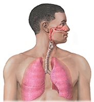
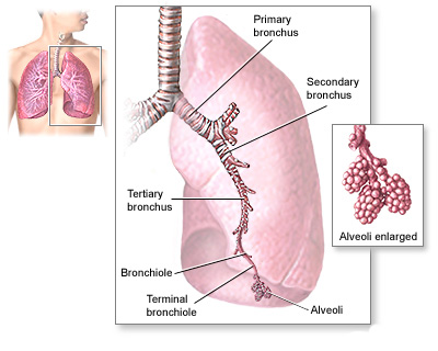
Breathing is a complex process. If injury, disease, or other factors affect any part of the process, you may have trouble breathing.
For example, the fine hairs (cilia) that line your upper airways may not trap all of the germs you breathe in. These germs can cause an infection in your bronchial tubes (bronchitis) or deep in your lungs (pneumonia). These infections cause a buildup of mucus or fluid that narrows the airways and limits airflow in and out of your lungs.
If you have asthma, breathing in certain substances that you're sensitive to can trigger your airways to narrow. This makes it hard for air to flow in and out of your lungs.
Over a long period, breathing in cigarette smoke or air pollutants can damage the airways and air sacs. This can lead to a disease called COPD (chronic obstructive pulmonary disease). COPD prevents proper airflow in and out of your lungs and can hinder gas exchange in the air sacs.
An important step to breathing is the movement of your diaphragm and other muscles in your chest, neck, and abdomen. This movement lets you inhale and exhale. Nerves that run from your brain to these muscles control their movement. Damage to these nerves in your upper spinal cord can cause breathing to stop, unless a machine is used to help you breathe. (This machine is called a ventilatoror a respirator.)
A steady flow of blood in the small blood vessels that surround your air sacs is vital for gas exchange. Long periods of inactivity or surgery can cause a blood clot called a pulmonary embolism (PE) to block a lung artery. A PE can reduce or block the flow of blood in the small blood vessels and hinder gas exchange.
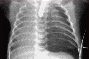
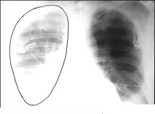
A collapsed lung, or pneumothorax, is the collection of air in the space around the lungs. This buildup of air puts pressure on the lung, so it cannot expand as much as it normally does when you take a breath.
A collapsed lung occurs when air escapes from the lung and fills up the space outside of the lung, inside the chest. It may be caused by a gunshot or knife wound to the chest, rib fracture, or certain medical procedures.
In some cases, a collapsed lung occurs without any cause. This is called a spontaneous pneumothorax. A small area in the lung that is filled with air (bleb) can break open, sending air into the space around the lung.
Tall, thin people and smokers are more likely to have a collapsed lung.
The following lung diseases also increase your risk for a collapsed lung:
A small pneumothorax may go away on its own. You may only need oxygen and rest.
The doctor may use a needle to pull the extra air out from around the lung so it can expand more fully. You may be allowed to go home if you live near the hospital.
If you have a large pneumothorax, a chest tube will be placed between the ribs into the space around the lungs to help drain the air and allow the lung to re-expand.
The chest tube can be left in place for several days. You may need to stay in the hospital. However, you may be able to go home if a small chest tube is used.
Some patients with a collapsed lung need extra oxygen.
Lung surgery may be needed to treat your pneumothorax or to prevent future episodes. The area where the leak occurred may be repaired. Sometimes, a special chemical is placed into the area of the collapsed lung. This chemical causes a scar to form. This procedure is called pleurodesis.