Osteonecrosis is bone death caused by poor blood supply. It is most common in the hip and shoulder, but can affect other large joints such as the knee, elbow, wrist and ankle.
Osteonecrosis occurs when part of the bone does not get blood and dies. After a while, the bone can collapse. If osteonecrosis is not treated, the joint deteriorates, leading to severe arthritis.
Osteonecrosis can be caused by disease or by severe trauma, such as a fracture or dislocation, that affects the blood supply to the bone. Osteonecrosis can also occur without trauma or disease. This is called idiopathic -- meaning it occurs without any known cause.
Your health care provider will do a physical exam to find out if you have any diseases or conditions that may affect your bones. You will be asked about your symptoms and medical history.
Be sure to let your provider know about any medicines or vitamin supplements you are taking, even over-the-counter medicine.
After the exam, your provider will order one or more of the following tests:
If your provider knows the cause of osteonecrosis, part of the treatment will be aimed at the underlying condition. For example, if a blood clotting disorder is the cause, treatment will consist, in part, of clot-dissolving medicine.
If the condition is caught early, you will take pain relievers and limit use of the affected area. This may include using crutches if your hip, knee, or ankle is affected. You may need to do range-of-motion exercises. Nonsurgical treatment can often slow the progression of osteonecrosis, but most people will need surgery.
Many cases of osteonecrosis do not have a known cause, so prevention may not be possible. In some cases, you can reduce your risk by doing the following:
· Avoid drinking excessive amounts of alcohol.
· When possible, avoid high doses and long-term use of corticosteroids.
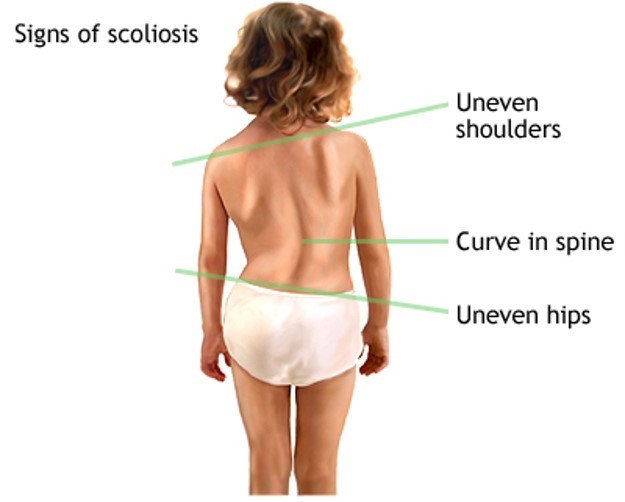
Scoliosis is an abnormal curving of the spine. Your spine is your backbone. It runs straight down your back. Everyone’s spine naturally curves a bit. But people with scoliosis have a spine that curves too much. The spine might look like the letter C or S.
Abnormal curvature in the spine is known as scoliosis, and generally begins just at the onset of puberty and progresses during the period of rapid growth. Most junior high schools routinely screen for scoliosis because, if caught early, progressive spine curvature can be prevented. Scoliosis affects girls much more frequently than boys.
Most of the time, the cause of scoliosis is unknown. This is called idiopathic scoliosis. It is the most common type. It is grouped by age.
In children age 3 and younger, it is called infantile scoliosis.
In children age 4 through 10, it is called juvenile scoliosis.
In children age 11 through 18, it is called adolescent scoliosis.
Scoliosis most often affects girls. Some people are more likely to have curving of the spine. Curving generally gets worse during a growth spurt.
Other types of scoliosis are:
Congenital scoliosis: This type of scoliosis is present at birth. It occurs when the baby’s ribs or spine bones do not form properly.
Neuromuscular scoliosis: This type is caused by a nervous system problem that affects the muscles. Problems can include cerebral palsy, muscular dystrophy, spina bifida, and polio.
The health care provider will perform a physical exam. You will be asked to bend forward. This makes your spine easier to see. It may be hard to see changes in the early stages of scoliosis.
The exam may show:
One shoulder is higher than the other
The pelvis is tilted
X-rays of the spine are done. X-rays are important because the actual curving of the spine may be worse than what your doctor can see during an exam.
Most people with idiopathic scoliosis do not need treatment. But you should still be checked by a doctor about every 6 months.
If you are still growing, your doctor might recommend a back brace. A back brace prevents further curving. There are many different types of braces. What kind you get depends on the size and location of your curve. Your health care provider will pick the best one for you and show you how to use it. Back braces can be adjusted as you grow.
Back braces work best in people over age 10. Braces do not work for those with congenital or neuromuscular scoliosis.
Scoliosis surgery involves correcting the curve as much as possible.
The spine bones are held in place with one or two metal rods. The rods are held down with hooks and screws until the bone heals together.
Surgery may be done with a cut through the back, belly area, or beneath the ribs.
After surgery, you may need to wear a brace for a while to keep the spine still.
You may need surgery if the spine curve is severe or getting worse very quickly. The surgeon may want to wait until all your bones stop growing, but this is not always possible.
Scoliosis treatment may also include:
Emotional support. Some children, especially teens, may be self-conscious when using a back brace.
Physical therapy and other specialists to help explain the treatments and make sure the brace fits correctly.
How well a person with scoliosis does depends on the type, cause, and severity of the curve. The more severe the curving, the more likely it will get worse after the child stops growing.
People with mild scoliosis do well with braces. They usually do not have long-term problems. Back pain may be more likely when the person gets older.
Outlook for those with neuromuscular or congenital scoliosis varies. They may have another serious disorder such as cerebral palsy or muscular dystrophy, so their goals are much different. Often the goal of surgery is simply to allow a child to be able to sit upright in a wheelchair.
Congenital scoliosis is difficult to treat and usually requires many surgeries.
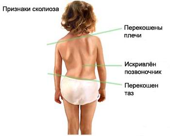
Сколиоз – аномальное искривление позвоночника. Ваш позвоночник – главная опора всего организма, расположенная в центре спины. У любого здорового человека позвоночник немножко изгибается. Однако люди, страдающие от сколиоза, имеют слишком сильный изгиб позвоночного столба. Изгиб позвоночника у таких людей иногда может напоминать букву «С» или латинскую «S».
Причины возникновения
В большинстве случаев причина появления сколиоза неизвестна. В таком случае сколиоз называют идиопатический сколиоз - это самый распространенный тип искривления позвоночника. Классифицируется это заболевание по возрастным группам.
У детей в возрасте до трех лет - инфантильный сколиоз.
У детей в возрасте 4-10 лет – ювенильный сколиоз
У детей постарше в возрасте 11-18 лет – подростковый сколиоз
Чаще всего от сколиоза страдают девочки. Некоторая категория людей анатомически имеет предрасположенность к сильному изгибу позвоночника. Обычно искривление позвоночника усиливается во время периода быстрого роста.
Другие типы сколиоза:
Врожденный сколиоз. Тип сколиоза, который выявляется сразу же после рождения ребенка. Возникает такая форма сколиоза в том случае, если кости ребер и позвоночника неправильно сформировались в молодом организме.
Нервно-мышечный сколиоз. Тип сколиоза, вызванный проблемами с нервной системой, которая влияет на мышцы. К примеру, церебральный паралич, мышечная дистрофия, расщепление позвоночника, и полиомиелит.
Симптомы
Обычно сколиоз проходит бессимптомно. В случае наличия, симптомы включают в себя:
Боль в спине и пояснице
Чувство усталости в позвоночнике после длительного нахождения в сидячем или стоячем положении
Неодинаковые пропорции бедер или плеч (одно плечо может быть выше другого).
Позвоночник сильнее искривляется в какую-либо сторону.
Еще одним типом аномального искривления позвоночника является кифосколиоз.
Диагностика
Для диагностики заболевания врач проводит физический осмотр. Также врач просит пациента наклониться вперед, что позволяет более подробно разглядеть позвоночник пациента. На ранних этапах развития сколиоза иногда изменения в позвоночнике бывает трудно разглядеть.
При осмотре может быть выявлено:
Что одно плечо выше другого
Таз наклонен в сторону
Для диагностики иногда необходимо сделать рентген позвоночника. Это важно, так как искривление может быть гораздо более серьезным, чем кажется при физическом осмотре.
В какой области находится искривление позвоночника
Насколько сильно искривление
Продолжает ли организм пациента расти
Большинство пациентов с идиопатическим сколиозом не требуют лечения. Тем не менее, таким пациентам необходимо наблюдаться у врача не менее одного раза в полгода.
Если организм пациента все еще растет, то врач может порекомендовать ношение корсета. Корсет устраняет дальнейшее искривление. Существует множество разнообразных корсетов, и они подбираются в зависимости от размера и области искривления позвоночника. Лечащий врач подбирает пациенту самый лучший корсет и показывает, как его использовать. Корсеты могут быть заменены по мере роста.
Корсеты помогают больше всего людям старше 10 лет. Однако корсеты неэффективны для лечения врожденного или нервно-мышечного сколиоза.
В некоторых случаях для лечения сколиоза требуется хирургическое вмешательство, направленное на устранение искривления. Во время операции кости позвоночника фиксируются специальными металлическими штифтами, которые держатся на крюках и болтах, пока кости полностью не срастутся. Операцию могут проводить, сделав разрез в области спины, живота, ребер.
После хирургического вмешательства пациенту необходимо носить корсет для фиксации позвоночника.
Хирургическое вмешательство также необходимо, если искривление очень сильное и ухудшается очень быстро. Хирург зачастую захочет дождаться момента, когда кости перестанут расти, чтобы провести операцию, но, к сожалению, это не всегда представляется возможным.
Лечение сколиоза также включает в себя:
Эмоциональную поддержку. Некоторые дети, особенно подростки, могут испытывать сильное чувство стеснения во время ношения корсета.
Физиотерапевты и другие специалисты могут объяснить цель лечения и убедиться, что корсет подходит пациенту.
Течение заболевания
Успешность устранения искривления позвоночника зависит от типа, причины и степени сколиоза. Чем сильнее искривлен позвоночник, тем сильнее вероятность того, что состояние позвоночника ухудшится, когда организм перестанет расти.
Люди, страдающие от легкой степени сколиоза, хорошо себя чувствуют в корсете. Обычно у людей с такой формой сколиоза не возникает долгосрочных проблем. Тем не менее, сколиоз увеличивает вероятность возникновения болей в спине в пожилом возрасте.
Результаты лечения пациентов с нервно-мышечным сколиозом, или врожденным сколиозом разнятся. Пациенты, страдающие от нервно-мышечного сколиоза, также имеют другое серьезное заболевание, поэтому способы лечения этого состояния отличаются. Зачастую цель хирургического вмешательства, в таком случае, - вернуть ребенку возможность нормально сидеть в инвалидном кресле.
Врожденный сколиоз очень тяжело лечить и обычно он требует неоднократных операций.
Возможные осложнения
Осложнения, появляющиеся при сколиозе, включают в себя:
Проблемы с дыханием (при тяжелой форме сколиоза)
Боль в пояснице
Низкая самооценка
Постоянные боли из-за износа костей позвоночника
Послеоперационная инфекция позвоночника
Повреждение позвоночника или нерва из-за неисправленного искривления, или операции на позвоночнике
Когда обращаться к врачу
Обратитесь к специалисту, если у вас есть подозрения, что ваш ребенок имеет искривление позвоночника.
Профилактика
Обследование на факт выявления сколиоза проводится систематично среди школьников средних и старших классов.
Что это такое? Остеонекроз иначе называют аваскулярным некрозом. Это гибель костной ткани в результате уменьшения или прекращения кровоснабжения костной ткани.
Что является причиной? Аваскулярный некроз может быть вызван остеомиелитом - воспалением костной ткани, травмой (переломом, смещением кости), а также радиационной болезнью. К аваскулярному некрозу могут привести такие заболевания как серповидно-клеточная анемия, подагра и системная красная волчанка. Длительное употребление алкоголя или лекарственных средств (например, кортикостероидов) повышает риск аваскулярного некроза.
К симптомам аваскулярного некроза относятся слабая, умеренная боль в месте поражения - бедре или паху, нарушения движения и хромота. Боль может быть резкой и обостряется в стоячем положении или при ходьбе. Обычно отдых облегчает боль. У детей, страдающих аваскулярным некрозом, могут наблюдаться спазмы в бедренной мышце, хромота или отказ от ношения тяжестей.
Обратитесь к вашему врачу или хирургу по месту жительства для того, чтобы узнать что следует предпринять.