The breast is made up of glands called lobules that can make milk and thin tubes called ducts that carry the milk from the lobules to the nipple. Breast tissue also contains fat and connective tissue, lymph nodes, and blood vessels.
Inside a woman’s breast are 15 to 20 sections (lobes). Each lobe is made of many smaller sections (lobules). Lobules have groups of tiny glands that can make milk. After a baby is born, breast milk flows from the lobules through thin tubes (ducts) to the nipple. Fibrous tissue and fat fill the spaces between the lobules and ducts.
Both males and females of all ages have normal breast tissue. This tissue responds to hormone changes. Because of this, lumps can come and go.
Breast lumps may appear at any age:
· Both male and female infants may have breast lumps from their mother's estrogen when they are born. The lump will most often go away on its own as the estrogen clears from the baby's body.
· Young girls often develop "breast buds," which appear just before the beginning of puberty. These bumps may be tender. They are common around age 9, but may happen as early as age 6.
· Teenage boys may develop breast enlargement and lumps because of hormone changes in mid-puberty. Although this may be upsetting to boys, the lumps or growth almost always go away on their own over a period of months.
Lumps in a woman are often caused by fibrocystic changes, fibroadenomas, and cysts.
Fibrocystic changes are painful, lumpy breasts. Fibrocystic breast changes do not increase your risk of breast cancer. Symptoms are most often worse right before your menstrual period, and then improve after your period starts.
Fibroadenomas are noncancerous lumps that feel rubbery. They move easily inside the breast tissue. Like fibrocystic changes, they occur most often during the reproductive years.They are most often not tender. Except in rare cases, they do not become cancerous later. A health care provider can feel during an exam whether a lump is a fibroadenoma. The only way to be sure, however, is to remove or biopsy the lump.
Cysts are fluid-filled sacs that often feel like soft grapes. These can sometimes be tender, often just before your menstrual period.
Other causes of breast lumps include:
· Breast cancer.
· Injury. Blood may collect and feel like a lump if your breast gets badly bruised. These lumps tend to get better on their own in a few days or weeks. If they do not improve, your provider may have to drain the blood.
· Lipoma. This is a collection of fatty tissue.
· Milk cysts (sacs filled with milk) and infections (mastitis), which may turn into an abscess. These typically occur if you are breastfeeding or have recently given birth.
See your provider if you have any new lumps or breast changes. Ask about your risk factors for breast cancer, and screening and prevention for breast cancer.
Your provider will get a complete history from you. You will be asked about your factors that may increase the risk of breast cancer. The provider will perform a thorough breast exam. If you don't know how to perform a breast self-exam, ask your provider to teach you the proper method.
You may be asked medical history questions such as:
· When and how did you first notice the lump?
· Do you have other symptoms such as pain, nipple discharge, or fever?
· Where is the lump located?
· Do you do breast self-exams, and is this lump a recent change?
· Have you had any type of injury to your breast?
· Are you taking any hormones, medicines, or supplements?
Steps your provider may take next include:
· Order a mammogram to look for cancer, or a breast ultrasound to see if the lump is solid or a cyst
· Use a needle to draw fluid out of a cyst, which will be examined under a microscope to look for cancer cells
How a breast lump is treated depends on the cause.
· Solid breast lumps are often removed with surgery.
· Cysts can be drained in the provider's office. If the fluid removed is clear or greenish, and the lump disappears after it is drained, you do not need further treatment. If the lump does not disappear or comes back, it is most often removed with surgery.
· Breast infections are treated with antibiotics.
· If you are diagnosed with breast cancer, you will discuss your options carefully and thoroughly with your provider.
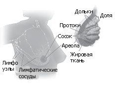
Молочная железа располагается на грудных мышцах, покрывающих ребра. Каждая молочная железа состоит из 15-20 долей. Доли содержат много мелких долек. Дольки состоят из групп крошечных желез, которые могут производить молоко. Молоко течет из долек по тонким трубочкам, которые называются протоками, к соску. Сосок расположен в центре темного круга кожи, называемого ареолой. Жировая ткань заполняет пространства между дольками и протоками.
В молочной железе также располагаются лимфатические сосуды. Эти сосуды ведут к маленьким округлым органам, называемым лимфатическими узлами. Группы лимфатических узлов локализуются рядом с молочной железой в подмышечной впадине, над ключицей, в грудной клетке за молочной железыной, а также во многих других местах тела. Лимфатические узлы задерживают бактерии, раковые клетки и другие вредные вещества.
Этот рисунок показывает строение молочной железы, а также лимфатические узлы и сосуды рядом с молочной железой.
Мастопатия (генерализированная комковатость молочной железы - фиброзно-кистозные изменения молочной железы) – это распространенное состояние молочной железы. Мастопатия - фиброзно-кистозная болезнь, для которой характерно изменение ткани молочной железы с нарушением соотношения соединительнотканного и эпителиального компонентов. Мастопатию диагностируют у 30-40% женщин с различными гормональными нарушениями (гиперэстрогения, прогестерондефицитное состояние и т.д.)
Генерализированная комковатость молочной железы обычно становится более заметной перед менструациями и ощущается в обеих грудях вокруг соска и в верхней наружной части грудей.
Генерализированная комковатость молочной железы наиболее часто встречается у женщин в возрасте между 35 и 50. Как правило, она исчезает с менопаузой. Она может продолжиться у женщин, принимающих эстроген для заместительной гормональной терапии.
Лечение мастопатии может назначаться только врачом после:
определения формы фиброзно-кистозного заболевания
обследования репродуктивной системы
определения гормонального статуса
Профилактика мастопатии заключается в своевременном обращении к врачу при выявлении изменений в груди. Важное значение имеет самообследование молочных желез, а также профилактические осмотры у маммолога женщинам после 45 лет.