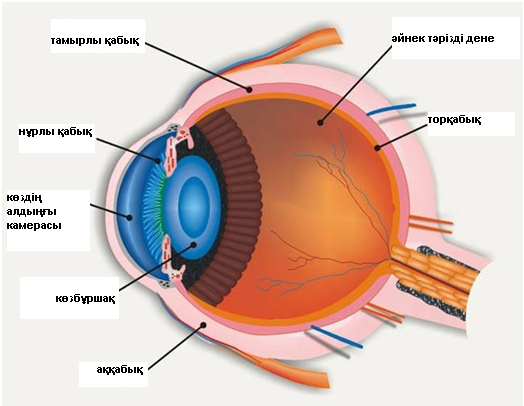
Көз ұясы үш қабықшадан және ішіндегілерден тұрады:
1) көздің сыртқы қабығы: аққабық (пішіні мен ширақтығын қамтамасыз ететін мөлдір емес бөлік), мөлдірқабық (жарық өтуі мен жарық сынуын қамтамасыз ететін мөлдір бөлік);
2) тамырлы қабық: нұрлы қабық (көзбұршақтың өлшемін реттейтін алдыңғы бөлігі), цилиарлық дене (көз ішілік сұйықтықты түзетін ортаңғы бөлік), хориоидея (торқабықты – көз түбін қоректендіретін артқы бөлік);
3) торқабық (өне бойымен ішкі жағымен әйнек тәрізді денеге жақын жататын көздің ішкі қабығы, ал сыртқы жағымен — тамырлы қабыққа жақын, алдыңғы көрмейтін бөліктен және біз көретін заттар бейнеленетін, артқы көретін бөліктен тұрады);
Көздің ішіндегісі: көзбұршақ (түрлі алыстағы заттарға көзді тоқтатуға-фокустеуге қатысатын биологиялық линза), әйнек тәрізді дене (мөлдір гель, көзбұршақты көз түбінен бөліп тұратын, көздің пішімін ұстап тұратын және жарық сәулесін торқабыққа өткізетін).
Торқабықтың ажырауы кенет пайда болып, көрмей қалуға апаруы мүмкін. Көбінесе, торқабықтың ажырауы орта және егде жастағы адамдарда пайда болады. алайда, бұл ауру балаларда, жаңа туған сәбилерде де болуы мүмкін.
· Алыстан көрмеудің орташа және жоғары дәрежесі (жақыннан жақсы көреді, алыстан нашар көреді) – бұл кезде көз ұясы ұзарады, торқабық созылады, шеттері жұқарады және үзіледі.
· Көздің жарақаты немесе офтальмологиялық операциялар.
· Қант диабетінде, қандағы қанттың деңгейі жоғары. Бұл қантамырларының зақымдануына апарады, оның ішінде, торқабықтың ұсақ тамырларының. Тамырлар жұқарады және жарылады, қан торқабықтың алдындағы бөлікке түседі, бұл көрудің бұзылуымен қосарланады. Қанның жиналуы тыртықтың түзілуіне апарады, олар торқабықты өзімен тартады, нәтижесінде, ол көздің тамырлы қабығынан ажырайды.
Торқабықтың ажырауы тек оперативтік тәсілмен емделеді.
· Криопексия (суықпен әсер ету) торқабықтың тұтастығын аурудың әр түрлі сатысында қалпына келтіруге қабілетті.
· Лазермен емдеу көздің торлы және тамырлы қабығының арасында бірігу түзілуі үшін қолданылады.
· Пневматикалық ретинопексия — хирургиялық емдеу тәсілі, ажыраудың кейбір түрінде тиімді. Бұл кезде әйнек тәрізді денеге көз ұясының ішіне ауа көпіршігін енгізеді, ол кеңейе отырып, торқабықты көздің қабырғасына қысады. Бұл шарадан кейін қосымша лазерлік емдеу қажет болады. 85% жағдайда пневматикалық ретинопексия көруді қалпына келтіреді.
· Склералау – склералық таңғыш аталатын арнайы силикон лентаны көз ұясын айналдыра орналастыру, бұл торқабықты «жабыстыруға» мүмкіндік береді. Склералық таңғыш (көздегі бандаж) торқабықты тамырлы қабыққа тығыздап, қысады және көрінбейді. Склералық лентаны орнатқаннан кейін оны қыздырумен жалғайды. Торқабықты склералаудың тиімділігі - 95% жуық.
· Витрэктомия – шағын тіліктер салу, олар арқылы әйнек тәрізді дене алынып, арнайы ерітінділермен алмастырылады. Витрэктомияның тиімділігі склералаудың тиімділігіне жуық.
Емдеудің табыстылығы торқабықты ажыраған тұсының орналасуына және емдеудің уақытылы болуына байланысты. Егер көздің макуласы (көрудің фокустелуіне жауап беретін бөлік) зақымданбаған болса, операциядан кейін көруді қалпына келтіру мүмкіндігі зор. Торқабықтың ажырауының көптеген түрлері емделеді, бәрі болмаса да. Операциядан кейін көру ішінара ғана қалпына келуі мүмкін.
· Күннен қорғайтын көзілдірік, көз жарақатын болдырмау үшін.
· Диабет кезінде қандағы қант деңгейін бақылау.
· Офтальмологқа бар, кемінде, жылына бір рет.
Retinal detachment is a separation of the light-sensitive membrane in the back of the eye (the retina) from its supporting layers.
CAUSES.
The most common type of retinal detachments are often due to a tear or hole in the retina. Eye fluids may leak through this opening. This causes the retina to separate from the underlying tissues, much like a bubble under wallpaper. This is most often caused by a condition called posterior vitreous detachment. However, it may also be caused by trauma and very bad nearsightedness. A family history of retinal detachment also increases your risk.
Another type of retinal detachment is called tractional detachment. This is seen in people who have uncontrolled diabetes, previous retinal surgery, or have chronic inflammation.
When the retina becomes detached, bleeding from area blood vessels may cloud the inside of the eye, which is normally filled with vitreous fluid. Central vision becomes severely affected if the macula, the part of the retina responsible for fine vision, becomes detached.
SYMPTOMS
Bright flashes of light, especially in peripheral vision
Most people with a retinal detachment will need surgery. Surgery may be done immediately or after a short period of time.
Surgery may not be needed if you do not have symptoms or have had the detachment for a while.
Some types of retinal detachment surgery can be done in your doctor's office.
Lasers may be used to seal tears or holes in the retina before a retinal detachment occurs.
If you have a small retinal detachment, the doctor may place a gas bubble in the eye. This is called pneumatic retinopexy. It helps help the retina float back into place. The hole is sealed with a laser.
More severe detachments may require surgery in a hospital operating room. Such procedures include:
Scleral buckle to gently push the eye wall up against the retina
Vitrectomy to remove gel or scar tissue pulling on the retina, used for the largest tears and detachments.
Tractional retinal detachments may be watched for a while before surgery. If surgery is needed, a vitrectomy is usually done.
PROGNOSIS
How well you do after a retinal detachment depends on the location and extent of the detachment and early treatment. If the macula was not damaged, the outlook with treatment can be excellent.
Most retinal detachments can be repaired, but not all of them. You may not get back all of your vision after surgery.
POSSIBLE COMPLICATIONS
A retinal detachment causes loss of vision. Surgery to repair it may help restore some or all of your vision.
WHEN TO CONTACT ВРАЧА
A retinal detachment is an urgent problem that requires medical attention within 24 hours of the first symptoms.
PREVENTION
Use protective eye wear to prevent eye trauma. Control your blood sugar carefully if you have diabetes. See your eye care specialist at least yearly, especially if you have risk factors for retinal detachment.
Dry eye usually occurs in people who are otherwise healthy. It becomes more common with age. This can occur due to hormonal changes that make your eyes produce fewer tears.
Other common causes of dry eyes include:
Dry environment or workplace (wind, air conditioning)
The first step in treatment is artificial tears. These come as preserved (screw cap bottle) and unpreserved (twist open vial). Preserved tears are more bottle) convenient, but some people are sensitive to preservatives. There are many brands available without a prescription.
Start using the drops at least 2-4 times per day. If your symptoms are not better after a couple of weeks of regular use:
Increase use (up to every 2 hours)
Try a different brand
Talk to your health care provider if you can’t find a brand that works for you
Other medical treatments may include:
Fish oil 2-3 times per day
Glasses, goggles or contact lenses that keep moisture in the eyes
Medicines such as Restasis, topical corticosteroids, and oral tetracycline and doxycycline
Tiny plugs placed in the tear drainage ducts to moisture stay on the surface of the eye longer
Don't smoke and avoid second-hand smoke, direct wind, and air conditioning.
Use a humidifier, especially in the winter.
Limit allergy and cold medicines that may dry you out and worsen your symptoms.
Purposefully blink more often. Rest your eyes once in a while.
Clean eyelashes regularly and apply and warm compresses.
Some dry eye symptoms are due to sleeping with the eyes slightly open. Lubricating ointments work best for this problem. You should use them only in small amounts since they can blur your vision. It is best to use them before sleep.
Surgery may be helpful if symptoms are because the eyelids are in an abnormal position.
The conjunctiva is exposed to bacteria and other irritants. Tears help protect the conjunctiva by washing away bacteria. Tears also contain proteins and antibodies that kill bacteria.
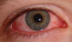
There are many causes of conjunctivitis. Viruses are the most common cause. Viral conjuctivitis is referred to as "pink eye." Pink eye can spread easily among children.
Use of contact lenses (especially extended-wear lenses)
Newborns can be infected by bacteria in the birth canal. This condition is called ophthalmia neonatorum, and it must be treated immediately to preserve eyesight.
Allergic conjunctivitis may respond to allergy treatment. It may disappear on its own when you avoid your allergy triggers. Cool compresses may help soothe allergic conjunctivitis.
Antibiotic medication, usually eye drops, is effective for bacterial conjunctivitis. Viral conjunctivitis will disappear on its own. Many doctors give a mild antibiotic eyedrops for pink eye to prevent bacterial conjunctivitis.
You can soothe the discomfort of viral or bacterial conjunctivitis by applying warm compresses (clean cloths soaked in warm water) to your closed eyes.