The eye will often flush out small objects, like eyelashes and sand, through blinking and tearing. DO NOT rub the eye if there is something in it. Wash your hands before examining the eye.
Examine the eye in a well-lighted area. To find the object, look up and down, then from side to side.
· If you can't find the object, grasp the lower eyelid and gently pull down on it to look inside the lower eyelid. To look inside the upper lid, you can place a cotton-tipped swab on the outside of the upper lid and gently fold the lid over the cotton swab.
· If the object is on an eyelid, try to gently flush it out with water or eye drops. If that does not work, try touching a second cotton-tipped swab to the object to remove it.
· If the object is on the white of the eye, try gently rinsing the eye with water or eye drops. Or you can GENTLY touch a cotton swap to the object to try to remove it. If the object is on the colored part of the eye, DO NOT attempt to remove it. Your eye may still feel scratchy or uncomfortable after removing an eyelashes or other tiny object. This should go away within a day or two. If you continue to have discomfort or blurred vision, get medical help.
Contact your health care provider and DO NOT treat yourself if:
· You have a lot of eye pain or sensitivity to light.
· Your vision is decreased.
· You have red or painful eyes.
· You have flaking, discharge, or a sore on your eye or eyelid.
· You have had trauma to your eye, or you have a bulging eye or a drooping eyelid.
· Your dry eyes do not get better with self-care measures within a few days.
If you have been hammering, grinding, or could have come in contact with metal fragments, DO NOT attempt any removal. Go to the nearest emergency room immediately.
Presbyopia is a condition in which the lens of the eye loses its ability to focus, making it difficult to see objects up close.
CAUSES
In the young eye, the lens needs to change its length or shape to focus on objects that are close. The ability of the lens to change shape is called the elasticity of the lens. This elasticity is slowly lost as people age. The result is a slow decrease in the ability of the eye to focus on nearby objects.
People usually notice the condition at around age 45, when they realize that they need to hold reading materials further away in order to focus on them. Presbyopia is a natural part of the aging process and it affects everyone.
SYMPTOMS
Decreased focusing ability for near objects
Eyestrain
Headache
EXAMS AND TESTS
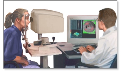
The health care provider will perform a general eye examination, including measurements to determine a prescription for glasses or contact lenses.
There is no cure for presbyopia, but it can be corrected with glasses or contact lenses. In some cases, adding bifocals to an existing lens prescription is enough. As the ability to focus up close worsens, the bifocal prescription needs to be strengthened.
Around age 65, the eyes have usually lost most of the elasticity needed to focus up close. However, it will still be possible to read with the help of the right prescription. Even so, you may find that you need to hold reading materials farther away, and you may need larger print and more light by which to read.
People who do not need glasses for distance vision may only need half glasses or reading glasses.
People who are nearsighted may be able to take off their distance glasses to read.
With the use of contact lenses, some people choose to correct one eye for near and one eye for far vision. This is called "monovision," and it eliminates the need for bifocals or reading glasses, but it can affect depth perception.
Sometimes monovision can be produced through laser vision correction. There are also bifocal contact lenses that can correct for both near and far vision in both eyes.
New surgical procedures can also provide solutions for people who do not want to wear glasses or contacts.
OUTLOOK (PROGNOSIS)
Vision can be corrected with glasses or contact lenses.
POSSIBLE COMPLICATIONS
If it is not corrected, vision difficulty that gets worse over time can cause problems with driving, lifestyle, or work.
WHEN TO CONTACT A MEDICAL PROFESSIONAL
Call your health care provider or ophthalmologist if you have eye strain or are less able to focus on close objects.
Macular degeneration is an eye disorder that slowly destroys sharp, central vision. This makes it difficult to see fine details and read.
The disease is most common in people over age 60, which is why it is often called age-related macular degeneration (ARMD, or AMD).
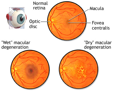
Macular degeneration is a disease of the retina that affects the macula in the back of the eye. The macula is important for clear central vision, allowing an individual to see fine details. There are two types of macular degeneration, dry and wet. Dry macular degeneration is more common and is characterized by the thinning of the retina and drusen, small yellowish-white deposits that form within the retina. The dry form of macular degeneration is usually mild. Wet macular degeneration can happen more quickly and be more serious. It occurs when vessels under the retinal layer hemorrhage and cause the retinal cells to die creating blind spots or distorted vision in the central vision. The disease becomes increasingly common amongst people in each succeeding decade over 50.
The retina is at the back of the eye. It changes light and images that enter the eye into nerve signals that are sent to the brain. A part of the retina called the macula makes vision sharper and more detailed.
AMD is caused by damage to the blood vessels that supply the macula. This change also harms the macula.
There are two types of AMD:
Dry AMD occurs when the blood vessels under the macula become thin and brittle. Small yellow deposits, called drusen, form. Almost all people with macular degeneration start with the dry form.
Wet AMD occurs in about 10% of people with macular degeneration. New abnormal and very fragile blood vessels grow under the macula. These vessels leak blood and fluid. This type of AMD causes most of the vision loss associated with the condition.
Doctors are not sure what causes AMD. The condition is rare before age 55. It occurs most in persons 75 years or older.
You may not have any symptoms at first. As the disease gets worse, you may have problems with your central vision.
SYMPTOMS OF DRY AMD
The most common symptom of dry AMD is blurred vision. Objects in the center part of your vision often look distorted and dim, and colors look faded. You may have trouble reading print or seeing other details. But you can see well enough to walk and do most daily activities.
As dry AMD gets worse, you may need more light to read or do everyday tasks. A blurred spot in the center of vision gradually gets larger and darker.
In the later stages of dry AMD, you may not be able to recognize faces until they are close.
SYMPTOMS OF WET AMD
The most common early symptom of wet AMD is that straight lines look distorted and wavy.
There may be a small dark spot in the center of your vision that gets larger over time.
With both types of AMD, central vision loss can occur quickly. If this happens, you will need to be seen right away by an ophthalmologist. Make sure this eye doctor has experience in treating problems with the retina.
You will have an eye exam. Drops will be placed into your eyes to widen (dilate) your pupils. The eye doctor will use special lenses to view your retina, blood vessels, and optic nerve.
The eye doctor will look for specific changes in the macula and blood vessels and for drusen.
You may be asked to cover one eye and look at a pattern of lines called an Amsler grid. If the straight lines look wavy, it may be a sign of AMD.
Other tests that may be done include:
Using special dye and camera to look at blood flow in the retina (fluorescein angiogram)
Taking a photo of the inner lining of the eye (fundus photography)
Using light waves to view the retina (optical coherence tomography)
If you have advanced or severe dry AMD, no treatment can restore your vision.
If you have early AMD and do not smoke, a combination of certain vitamins, antioxidants, and zinc may prevent the disease from getting worse. But it cannot give you back vision that is already lost.
The combination is often called the "AREDS" formula. The supplements contain:
500 milligrams of vitamin C
400 international units of beta-carotene
80 milligrams of zinc
2 milligrams of copper
Only take this vitamin combination if your doctor recommends it. Make sure your doctor knows about any other vitamins or supplements you are taking. Smokers should not use this supplement.
AREDS may also benefit you if you have a family history and risk factors for AMD.
The supplements lutein and zeaxanthin may also be helpful, although they are not part of the AREDS formula.
If you have wet AMD, your doctor may recommend:
Laser surgery (laser photocoagulation) -- a small beam of light destroys the leaking, abnormal blood vessels.
Photodynamic therapy -- a light activates a drug that is injected into your body to destroy leaking blood vessels.
Special medicines that prevent new blood vessels from forming in the eye are injected into the eye (this is a painless process).
Low-vision aids (such as special lenses) and therapy can help you use the vision that you have more effectively, and improve your quality of life.
Close follow-up with your eye doctor is important.
For dry AMD, visit your eye doctor once a year for a complete eye exam.
For wet AMD, you likely need frequent, perhaps monthly, follow-up visits.
Early detection of vision changes is important because the sooner you are treated, the better your outcome. Early detection leads to earlier treatment and often, a better outcome.
The best way to detect changes is by self-testing at home with the Amsler grid. Your eye doctor can give you a copy of the grid. Or you can print one from the Internet. Test each eye individually while wearing your reading glasses. If the lines look wavy, call your eye doctor right away for an appointment.
AMD does not affect side (peripheral) vision. This means complete vision loss never occurs. AMD results in the loss of central vision only.
Mild, dry AMD usually does not cause disabling central vision loss.
Wet AMD often leads to significant vision loss.
In general, with AMD you may lose the ability to read, drive a car, and recognize faces at a distance. But most people with AMD can carry out daily tasks without much difficulty.
If you have AMD, your health care provider may recommend that you check your vision every day with an Amsler grid. Call your provider immediately if the lines look wavy. Also call if you notice other changes in your vision.
Fuchs' (pronounced Fooks) dystrophy is an eye disease in which cells lining the inner surface of the cornea slowly start to die off. The disease usually affects both eyes.
Fuchs' dystrophy can be inherited, which means it can be passed down from parents to children. If either of your parents has the disease, you have a 50% chance of developing the condition.
However, the condition may also occur in persons without a known family history of the disease.
Fuchs' dystrophy is more common in women than in men. Vision problems usually do not appear before age 50, although doctors may be able to see signs of the disease in affected persons at an earlier age, usually in their 30s and 40s.
Fuchs' dystrophy affects the thin layer of cells that line the back part of the cornea. These cells help pump excess fluid out of the cornea. As more and more cells are lost, fluid begins to build up in the cornea, causing swelling and a cloudy cornea.
At first, fluid may build up only during sleep, when the eye is closed. As the disease gets worse, small blisters may form. The blisters get bigger and may eventually break, causing eye pain. Fuchs' dystrophy can also cause the shape of the cornea to change, causing further vision problems.
Deep lamellar keratoplasty (DLK) is an alternative to a traditional transplant. In this procedure, only the deep layers of the cornea are replaced with donor tissue. The procedure requires no stitches. Recovery time is faster and there are fewer complications, such as rejection.
Fuchs' dystrophy gets worse over time. Without a corneal transplant, a patient with severe Fuchs' dystrophy may become blind or have severe pain and very reduced vision.
Mild cases of Fuchs' dystrophy often worsen after cataract surgery. A cataract surgeon will evaluate this risk and may modify the technique or the timing of your cataract surgery.
There is no known prevention. Avoiding cataract surgery or taking special precautions during cataract surgery may help slow down the course of the disease.
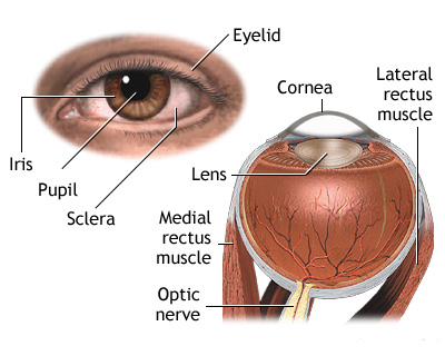
The most common form of uveitis is anterior uveitis, which involves inflammation in the front part of the eye. It is often called iritis because it usually only affects the iris, the colored part of the eye. The inflammation may be associated with autoimmune diseases, but most cases occur in healthy people. The disorder may affect only one eye. It is most common in young and middle-aged people.
Posterior uveitis affects the back part of the uvea, and involves primarily the choroid, a layer of blood vessels and connective tissue in the middle part of the eye. This type of uveitis is called choroiditis. If the retina is also involved, it is called chorioretinitis. You may develop this condition if you have had a body-wide (systemic) infection or if you have an autoimmune disease.
Another form of uveitis is pars planitis. This inflammation affects the narrowed area (pars plana) between the colored part of the eye (iris) and the choroid. Pars planitis usually occurs in young men and is generally not associated with any other disease. However, some evidence suggests it may be linked to Crohn's disease and possibly multiple sclerosis.
Uveitis can be associated with any of the following:
Pars planitis is often treated with steroid eye drops. Other medicines, including steroids taken by mouth, may be prescribed to help suppress the immune system.
Posterior uveitis treatment depends on the underlying cause but almost always includes steroids taken by mouth. Additional specialists in infectious disease or autoimmunity may be needed for such diseases as syphilis, tuberculosis, AIDS, sarcoidosis, or Behcet syndrome.
If the uveitis is caused by a body-wide infection, treatment may involve antibiotics and powerful anti-inflammatory medicines called corticosteroids. See autoimmune disorders for information on treating such diseases.
Call for an appointment with your health care provider if you have symptoms of uveitis. Eye pain or reduced vision are urgent symptoms that require prompt medical attention.